
Image
|
Figure Caption
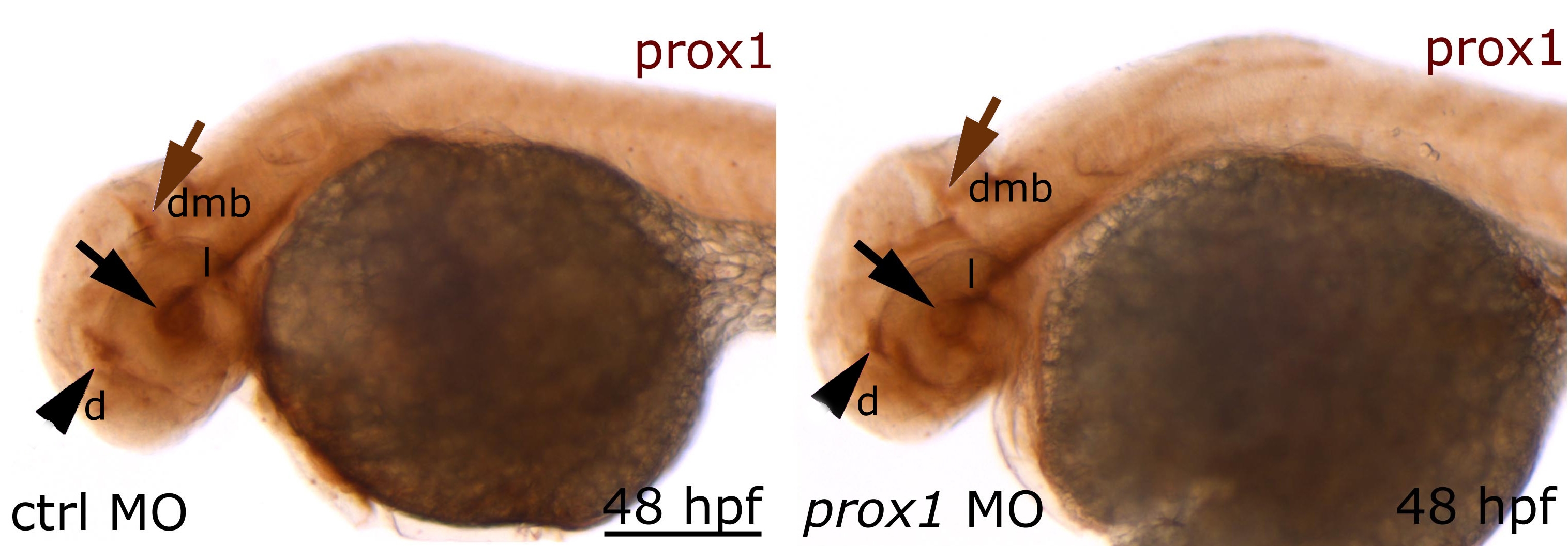
Fig. S1 Decreased levels of Prox1 protein in prox1 loss of function embryos. Immunohistochemistry using an anti-Prox1 antibody at 48 hpf (A) Prox1 protein distribution in control embryos in comparison to prox1 MO injected embryos (B) black arrow lens (l); arrowhead diencephalon (d), brown arrow diencephalic-mesencephalic boundary (dmb). Scale bar = 200 micron.
Figure Data
Acknowledgments
This image is the copyrighted work of the attributed author or publisher, and
ZFIN has permission only to display this image to its users.
Additional permissions should be obtained from the applicable author or publisher of the image.
Full text @ BMC Dev. Biol.