
|
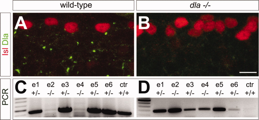
Fig. 2 Dla antibody is specific to zebrafish Dla protein. A and B show confocal images of lateral views of two spinal cord segments at 24 hours postfertilization (hpf); anterior to the left, dorsal up. Dla protein is revealed in green, Islet 1/2 (Isl) protein in red. A: Wild-type embryo showing punctate Dla labeling scattered throughout the spinal cord. B: Dla labeling is abolished in homozygous carriers in the viral insertion line dlahi781Tg (dla-/-). Isl labeling shows an increased number of RB neurons in homozygous dlahi781Tg embryos. C,D: Polymerase chain reaction (PCR) on genomic DNA extracted from 6 single embryos (e1-e6) and one control wild-type embryo (ctr). Two different primer sets reveal carriers of the viral insertion in the dla locus versus noncarriers. C: In DNA from heterozygous (+/-) carriers and noncarriers (+/+), dla-specific primers amplify a specific product with the expected size of 450 bp. D: A specific product of 270 bp is amplified in heterozygous (+/-) and homozygous (-/-) carriers whereas no product appears in wild-types (+/+). Scale bar = 8 μm in A,B.