
Image
|
Figure Caption
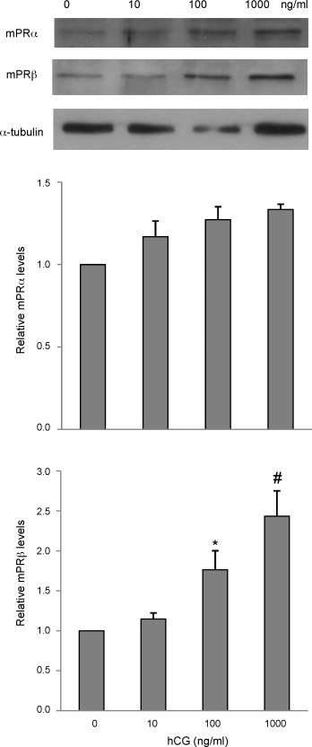
Fig. 4 Dose-dependent effect of hCG on mPR expression. Mid- to late growth phase follicles were treated with Cortland′s medium, or hCG at different concentrations (10, 100, and 1000 ng/ml) for 18 h. Proteins were extracted and expression of mPRα and mPRβ was detected by Western blot analysis. Equal loading was confirmed by Western blotting probed with an anti-α-tubulin antibody. Histograms show normalized densitometry data (mean ± S.E.M.) from three experiments. *p < 0.05 vs. control. #p < 0.05 vs. control and hCG 10 ng/ml groups.
Acknowledgments
This image is the copyrighted work of the attributed author or publisher, and
ZFIN has permission only to display this image to its users.
Additional permissions should be obtained from the applicable author or publisher of the image.
Reprinted from Molecular and Cellular Endocrinology, 312(1-2), Tan, Q., Zagrodny, A., Bernaudo, S., and Peng, C., Regulation of membrane progestin receptors in the zebrafish ovary by gonadotropin, activin, TGF-beta and BMP-15, 72-79, Copyright (2009) with permission from Elsevier. Full text @ Mol. Cell. Endocrinol.