
|
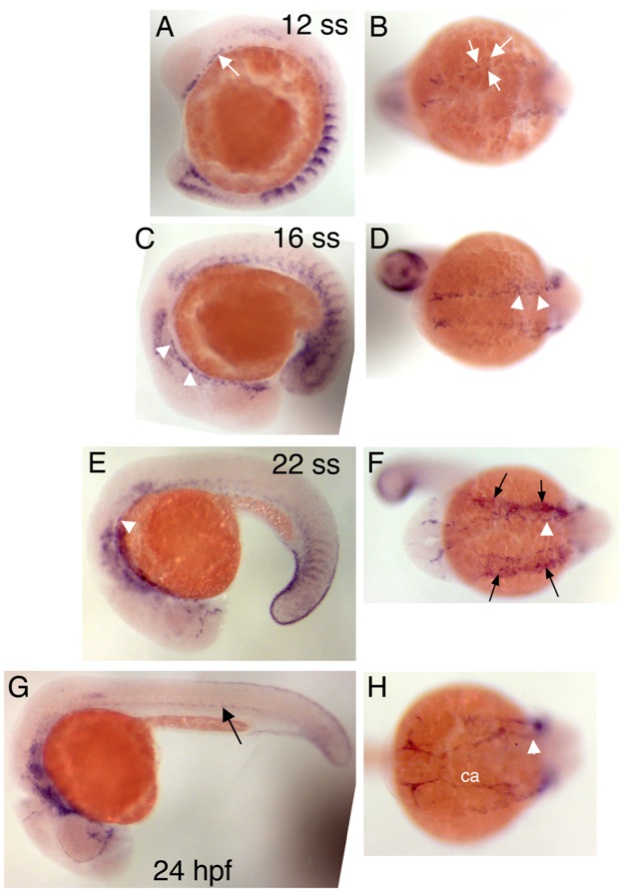
Fig. S3 Expression pattern of cxcr4a during zebrafish embryonic development. All embryos are shown with their anterior to the left, dorsal side up. A, C, E, G, side views, B, D, F, H, top views. A, B, 12 somite stage (ss;15 hours post fertilization), expression of cxcr4a can be detected in discrete cells in the anterior lateral mesoderm (arrows). C, D, 18 ss, anterior lateral dorsal aorta cells express cxcr4a (arrowheads). E, F, 22 ss, expression of cxcr4a continues in anterior lateral dorsal aorta cells (arrowheads). Strong expression of cxcr4a can also be detected in pharyngeal arch tissue (F, black arrows). G, H, 24 hpf, expression of cxcr4a can be detected in the lateral dorsal aorta (arrowhead), the head vasculature, for instance the carotid arteries (ca), and the dorsal aorta in the trunk (arrow).