
|
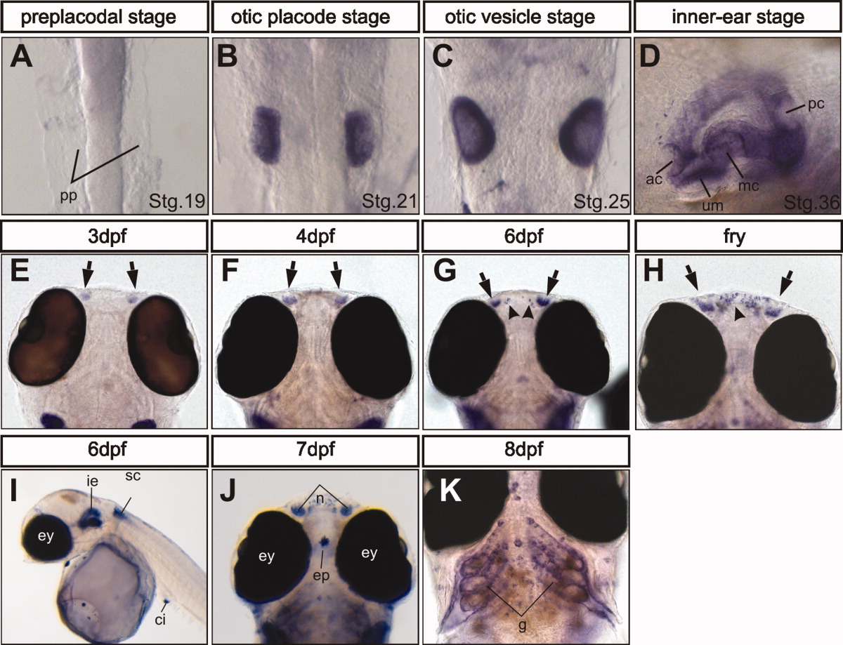
Fig. 2 Expression of the stm-l gene during embryonic development. Dorsal (A-C,E-H,J), ventral (K), and lateral (D,I) views of embryos. stm-l gene exhibits a specific expression in the otic structures (A-D) and nasal placode (E-H, arrows). The stm-l expression in the mouth region is detectable from 6 days postfertilization (dpf; G, arrowheads). stm-l is also expressed in the spinal cord (I), caudal intestine (I), epiphysis (J), and gills (K). Abbreviations: ac, anterior crista; ci, caudal intestine; ep, epiphysis; ey, eye; g, gills; ie, inner ear; mc, medial crista; n, nasal placode; pc, posterior cristae; pp, preplacodal region; sc, spinal cord; stg, stage; um, utricular macula.