
|
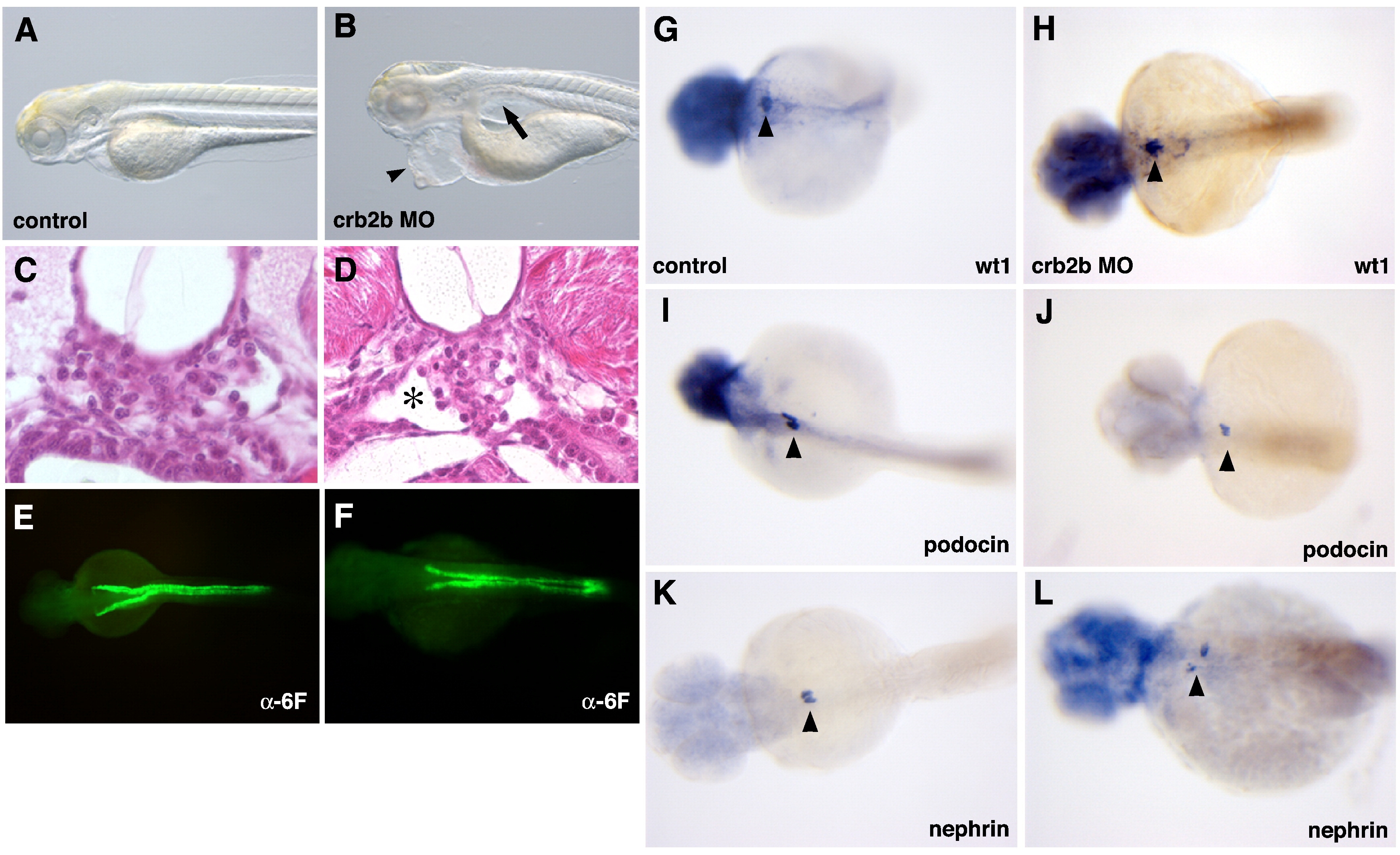
Fig. 3 Phenotype of the crb2b morphants. (A) Control mismatch and (B) crb2b-ATG injected 2.5 dpf larvae. Note the pericardial edema (arrowhead) and pronephric cysts (arrow). (C, D) Histological sections at the level of the glomerulus in control (C) and crb2b-ATG MO (D). Note the expanded Bowman′s space (asterisk) in D. (E, F) Staining with ±-Na+/K+ ATPase in the pronephric tubules and ducts is not generally affected in crb2b MO. (G–L) In situ hybridization on 2 dpf wildtype (G, I, K) or crb2b-ATG (H, J, L) with wt1 (G, H), podocin (I, J), or nephrin (K, L) antisense probes. Cells expressing wt1, nephrin, and podocin are present in crb2b morphants (black arrowheads).
Reprinted from Developmental Biology, 334(1), Ebarasi, L., He, L., Hultenby, K., Takemoto, M., Betsholtz, C., Tryggvason, K., and Majumdar, A., A reverse genetic screen in the zebrafish identifies crb2b as a regulator of the glomerular filtration barrier, 1-9, Copyright (2009) with permission from Elsevier. Full text @ Dev. Biol.