
Image
|
Figure Caption
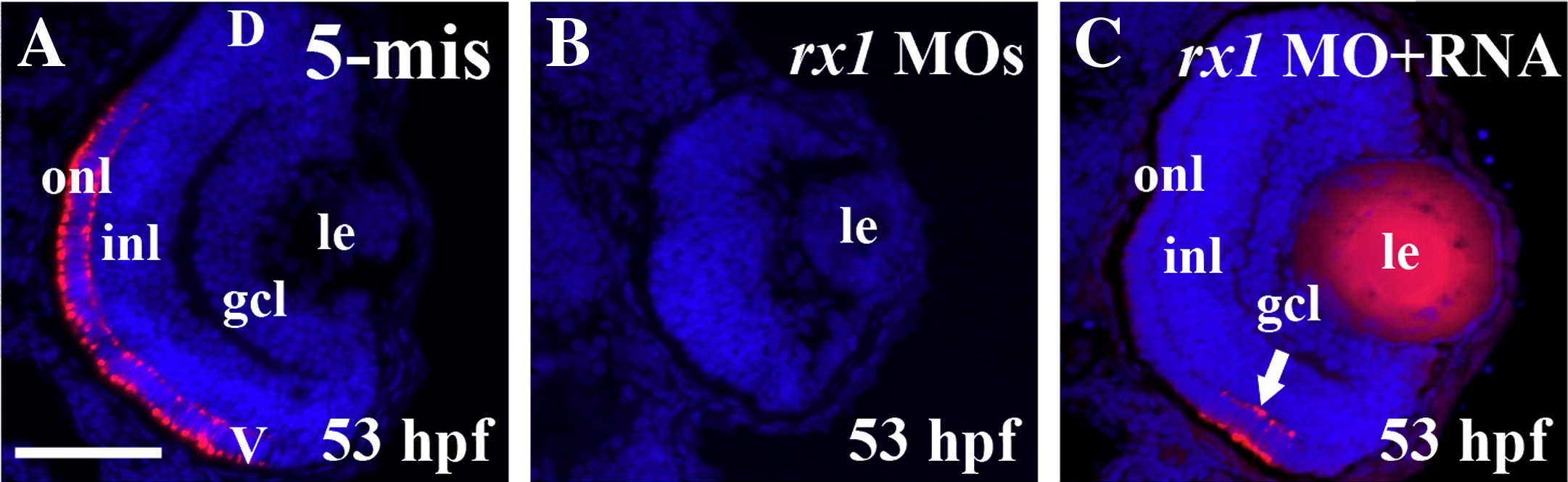
Fig. 2 Capped rx1 mRNAs rescue the rx1 depletion phenotype. (A–C) Sectioned embryos processed for indirect immunofluorescence with zpr1 (cone marker), and counterstained with DAPI to reveal retinal lamination. Embryos were injected with 5-mispair control MOs (A), splice site-directed rx1-MOs (B), or splice site-directed rx1-MOs in combination with capped rx1 mRNAs, arrow indicates zpr1+ photoreceptors in ventral retina (C). le = lens; gcl = ganglion cell layer; inl = inner nuclear layer; onl = outer nuclear layer; V = ventral; D = dorsal; scale bar in A (applies to all) = 50 μm.
Figure Data
Acknowledgments
This image is the copyrighted work of the attributed author or publisher, and
ZFIN has permission only to display this image to its users.
Additional permissions should be obtained from the applicable author or publisher of the image.
Reprinted from Developmental Biology, 328(1), Nelson, S.M., Park, L., and Stenkamp, D.L., Retinal homeobox 1 is required for retinal neurogenesis and photoreceptor differentiation in embryonic zebrafish, 24-39, Copyright (2009) with permission from Elsevier. Full text @ Dev. Biol.