
|
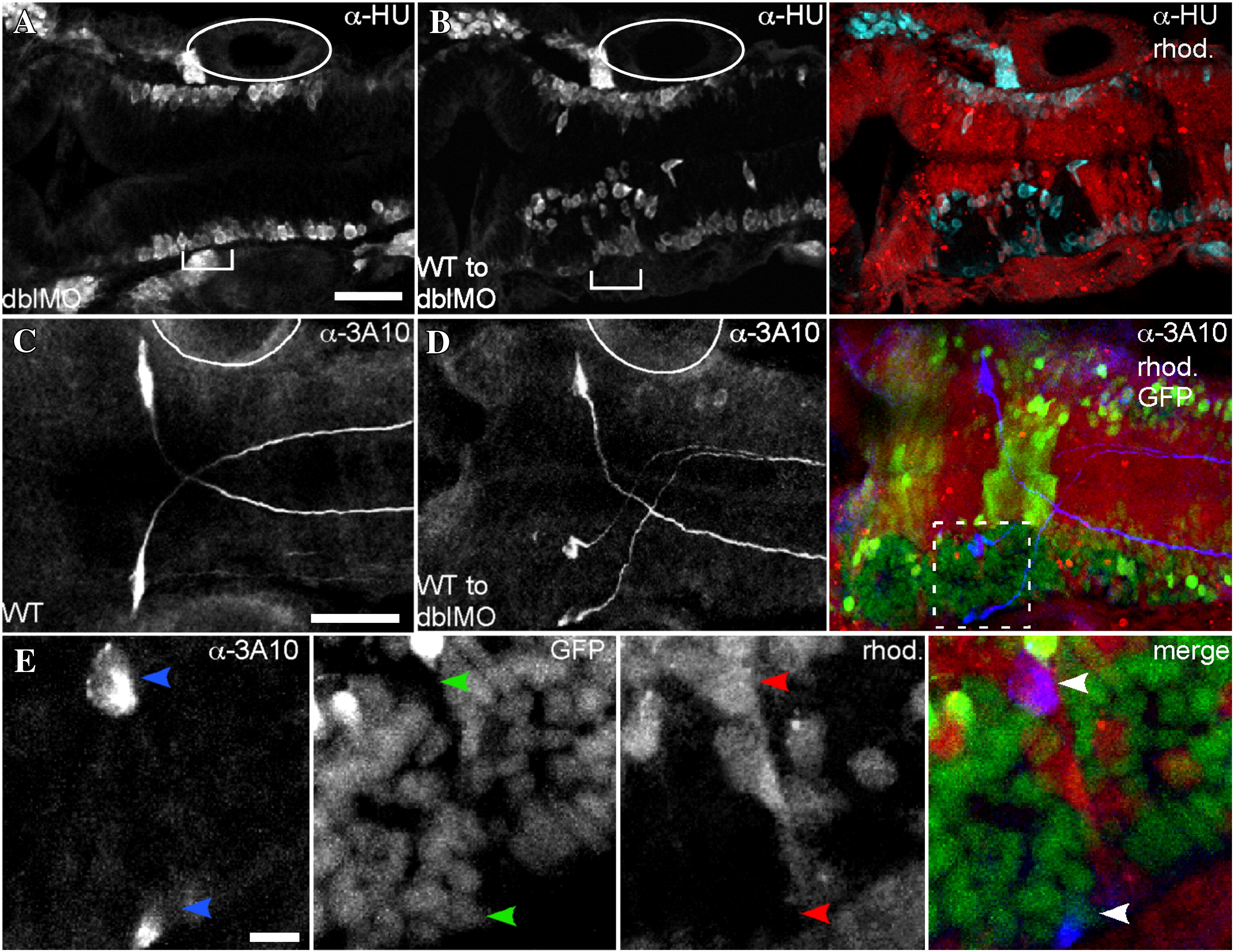
Fig. S8 WT cell clusters are neurogenic. Immunostaining of embryos shown in dorsal view with anterior to the left using α-HU at 26 hpf (A,B), or α-3A10 at 30 hpf (C–E). (A) In an EphA4;EfnB2a double MO embryo, neurogenesis, indicated by α-HU immunostaining, takes place at the lateral surfaces of the neuroepithelium. (B) Left panel shows α-HU immunostaining. Right panel is a merge showing rhodamine-injected double MO host cells surrounding unlabeled WT cell clusters. Ectopic neurogenesis is apparent at the edges of WT cell clusters (cyan). (C) α-3A10 shows the bilateral Mauthner neuron pair in r4 of a 30 hpf WT embryo. (D) Left panel shows α-3A10 immunostaining. Right panel is a merge of 3A10 staining in blue with rhodamine-injected double MO host cells (red) that also contain the noiGFP transgene (yellow) surrounding clusters of histone-GFP+ nuclei from WT donor cells (green). In WT to double MO mosaic embryos, ∼ 15% of embryos show duplication of the Mauthner neuron (N = 3/19). (E) Closeup view of the boxed region in D shows that one of the ectopic Mauthner neurons in r4 is donor-derived (histone-GFP+ nucleus, rhod.-negative cytoplasm). Scale bars: 50 μm (A–D), or 10 μm (E). Otic vesicle, adjacent to r5, is indicated by full or partial oval.
Reprinted from Developmental Biology, 327(2), Kemp, H.A., Cooke, J.E., and Moens, C.B., EphA4 and EfnB2a maintain rhombomere coherence by independently regulating intercalation of progenitor cells in the zebrafish neural keel, 313-326, Copyright (2009) with permission from Elsevier. Full text @ Dev. Biol.