
Fig. 3
|
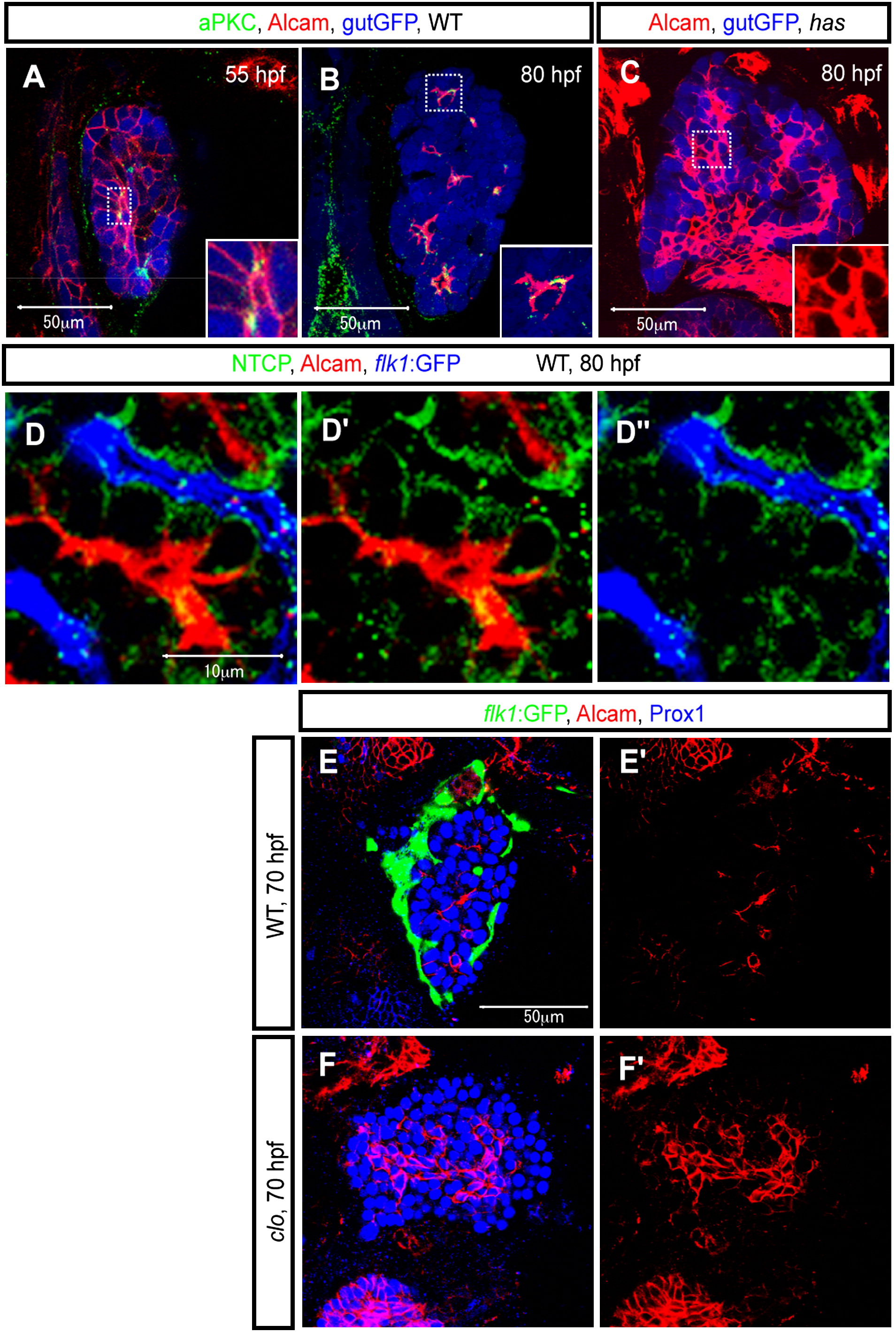
Fig. 3 Alcam Localizes to the Apical Membranes of Hepatocytes
Z-plane confocal images of the liver at 55 (A), 70 (E and F), and 80 (B–D) hpf. The images present ventral views, anterior to the top.
(A and B) Wild-type Tg(gutGFP)s854 larvae visualized for GFP (pseudocolored blue), Alcam (red), and aPKCs (pseudocolored green) expression. The outlined areas in (A) and (B) are magnified and shown in bottom right corners. At 55 hpf, aPKCs show restricted expression in hepatocytes prior to Alcam restriction. Alcam and aPKCs colocalize more extensively at 80 hpf.
(C) Tg(gutGFP)s854 heart and soulm567 (has) /prkci mutant larvae visualized for GFP (pseudocolored blue) and Alcam expression at 80 hpf. The Alcam staining of the outlined area is magnified and shown in the bottom right corner. At 80 hpf, whereas Alcam localization is restricted to the apical membrane of hepatocytes in wild-type larvae, in has m567 mutants, it is still localized along the entire cell membrane of most cells.
(D) Tg(flk1:EGFP)s843 larvae visualized for GFP (pseudocolored blue), Alcam (red), and Ntcp (green) expression. Alcam and Ntcp immunostainings are shown separately in (D′). Alcam and Tg(flk1:EGFP)s843 expressions are shown separately in (D″).
(E and F) Wild-type (E) and clochela1164 (clo) mutant (F) larvae visualized for Tg(flk1:EGFP)s843 (green), Alcam (red), and Prox1 (blue) expression at 70 hpf. At this stage, whereas Alcam has localized to the apical membranes of most cells in wild-type livers, it is still localized along the entire cell membrane of many cells in clo la1164 mutant livers.