
|
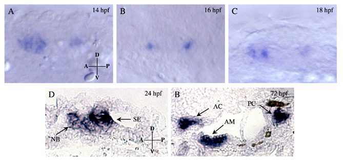
Fig. S1 eya4 expression during otic placode and otic vesicle development. (A-C) At 14, 16 and 18 hpf, wild-type embryos were hybridized to DIG-labeled eya4 antisense RNA probes. Lateral views of the otic placode region are shown. (D,E) Sections from wild-type embryos were hybridized with a DIG-labeled eya4 antisense RNA probe. eya4 signal (arrows) at 24 hpf (D) demarcated the developing sensory epithelia (SE) and neuroblast (NB). A section from 72 hpf (E) demonstrated eya4 expression in the anterior maculae (AM), anterior cristae (AC) and posterior cristae (PC) of the sensory epithelia. D, dorsal; V, ventral; A, anterior; P, posterior.