
|
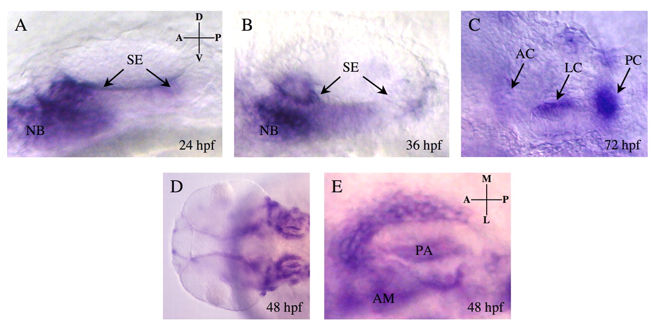
Fig. 1 eya4 expression during otic vesicle development. Wild-type embryos were hybridized to DIG-labeled eya4 antisense RNA probes. Lateral (A-C) and dorsal (E) views of single otic vesicles; (D) dorsal view of two otic vesicles. eya4 signal (arrows) at 24 hpf (A) and 36 hpf (B) demarcated the developing sensory epithelia (SE) and neuroblast (NB). At 72 hpf (C), expression was maintained in the anterior, lateral and posterior cristae (AC, LC and PC, respectively) of the sensory epithelia. At 48 hpf (D,E), eya4 signal was more diffuse within the otic vesicle and was prominent in the anterior macula (AM) and pharyngeal arch (PA; E). Background signal with eya4 sense RNA probe was minimal (data not shown). D, dorsal; V, ventral; A, anterior; P, posterior; M, medial; L, lateral.