
|
Fig. S2
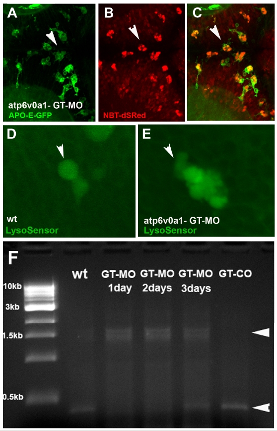
GT-MO injected embryos display a phenotype that is comparable with that observed for ATG-MO injected embryos
GT-MO morphant brain (A-C). APO-GFP (A), NBT.DsRed (B), merge (C). GT-MO injected embryos display a phenotype that is comparable with that observed for ATG-MO injected embryos (compare with Fig.4 L-O). Both are characterized an increase in the amount of NBT-DSRed neurons present in microglia (compare with Fig.4 H-K). D and E wild-type (D) and GT-MO (E) microglia stained with LysoSesor to label acidic vesicles. Intensity fluorescent values inside single vesicles (white arrowhead) are comparable, indicating normal acidification of these vesicles. (F) Efficacy of morpholino knock-down is confirmed by the 1.5kb size-shift in the transcript produced from the vAtpase gene (white arrowheads).
Reprinted from Cell, 133(5), Peri, F., and Nüsslein-Volhard, C., Live Imaging of Neuronal Degradation by Microglia Reveals a Role for v0-ATPase a1 in Phagosomal Fusion In Vivo, 916-927, Copyright (2008) with permission from Elsevier. Full text @ Cell