
|
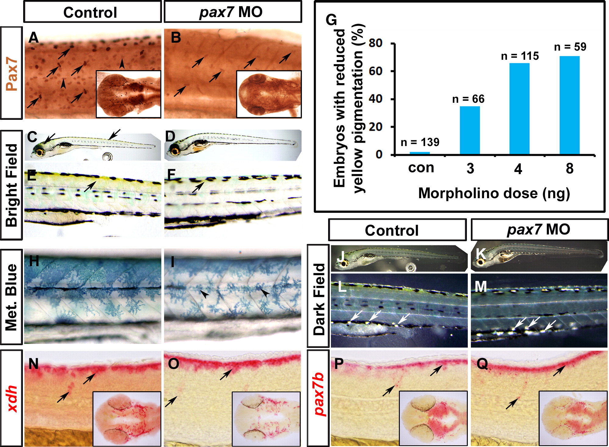
Fig. 6 Xanthophore pigmentation is reduced after knockdown of Pax7. Immunodetection of Pax7 (A, B), live images (C–F, H–M) and in situ mRNA hybridisation for xdh (N, O), pax7b (P, Q) of larvae. Lateral flatmounts are anterior to left and dorsal to top (A–F, H–Q) and dorsal flatmounts of head are anterior to left (insets, A, B, N–Q). (A, B) Pax7 protein is detected in migrating NC (arrows) and dermomyotome cells (arrowheads) at 26 hpf (A). Injection of a morpholino specific to pax7 results in ablation of Pax7 in dermomyotome cells and severely reduced reactivity in brain (inset) and NC (B, arrows). (C–F) At 6 dpf, yellow xanthophore pigmentation is present in the dorsal head and trunk (C, E; arrows). Pax7 morphants, show a marked reduction in yellow pigmentation in both head and trunk regions (D, F). (G) Dose-dependency of reduction in yellow pigmentation in pax7 morphants. (H, I) Methylene blue reveals morphological defects in pterinosome-containing cells after pax7 MO injection (I, arrowheads). (J–M) Dark field images at 6 dpf reveal iridophores in both control and pax7 morphants (arrows). (N–Q) xdh (N, O) and pax7b (P, Q) mRNAs persist in pax7 morphant xanthophore precursors (arrows).
Reprinted from Developmental Biology, 317(2), Minchin, J.E., and Hughes, S.M., Sequential actions of Pax3 and Pax7 drive xanthophore development in zebrafish neural crest, 508-522, Copyright (2008) with permission from Elsevier. Full text @ Dev. Biol.