
|
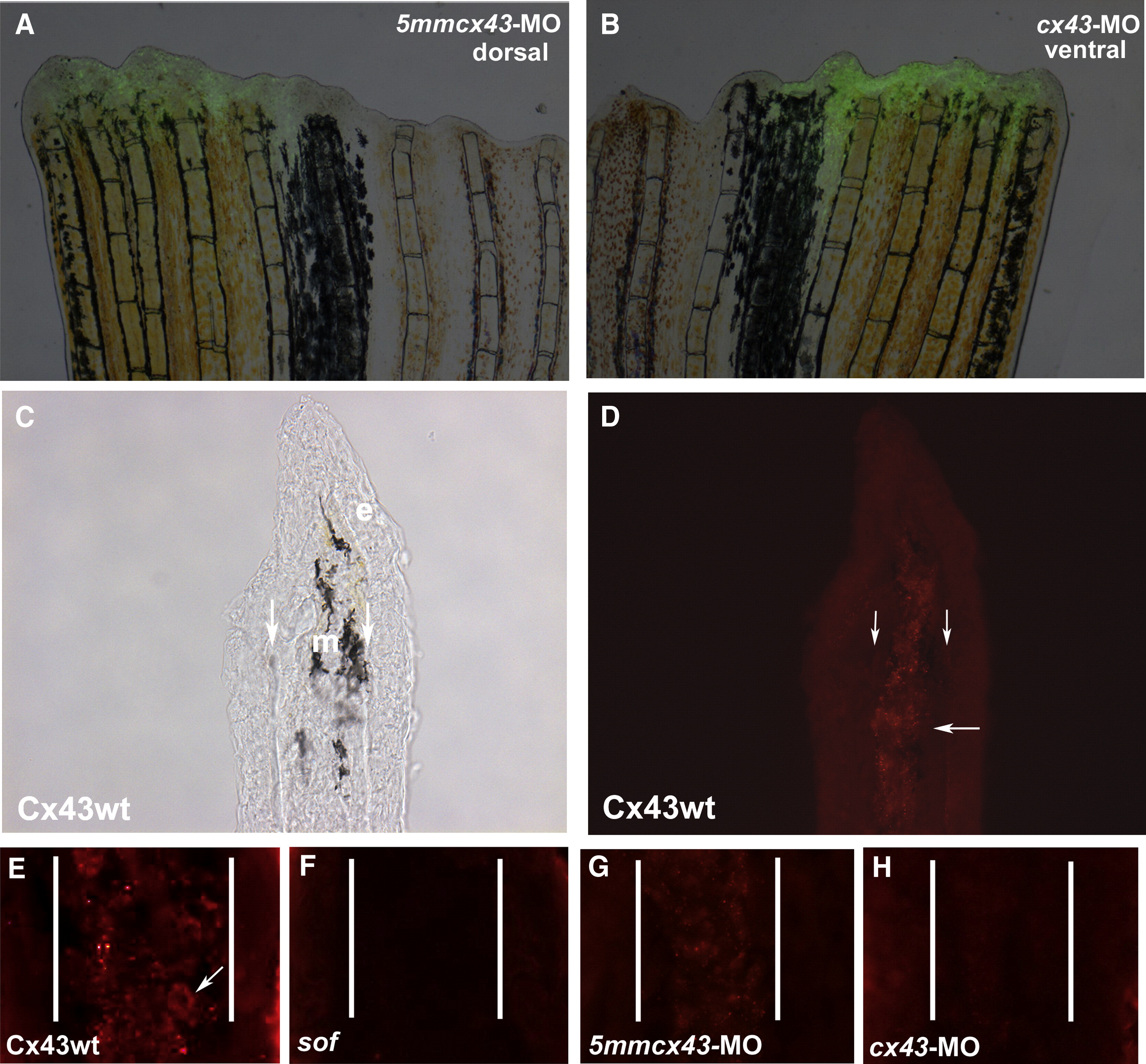
Fig. 5 Injection and electroporation of cx43 morpholinos confirms targeted knockdown of Cx43. (A,B) Fins were injected with either 1.2 mM 5mmcx43-MO or 1.2 mM cx43-MO and immediately electroporated. Cellular uptake is shown by fluorescein fluorescence. (C,D) Cryosections through fins stained with Cx43 antibody show mesenchymal staining in wild-type fins. Brightfield and fluorescence are shown. Vertical arrows indicate bone matrix, horizontal arrow points to one area of punctate staining. (E) Closer examination of Cx43 staining reveals punctate plasma membrane staining. Arrow points to one cell. (F) sofb123 regenerates exhibit a reduction in Cx43 protein in the mesenchyme (G) Fins injected/electroporated with the 5mmcx43-MO show similar expression of Cx43 as wild-type fins. (H) Fins injected/electroporated with the targeting morpholino cx43-MO exhibit a reduction in Cx43 expression. Vertical lines in panels E–H distinguish epithelium and mesenchymal compartments.
Reprinted from Developmental Biology, 317(2), Hoptak-Solga, A.D., Nielsen, S., Jain, I., Thummel, R., Hyde, D.R., and Iovine, M.K., Connexin43 (GJA1) is required in the population of dividing cells during fin regeneration, 541-548, Copyright (2008) with permission from Elsevier. Full text @ Dev. Biol.