
|
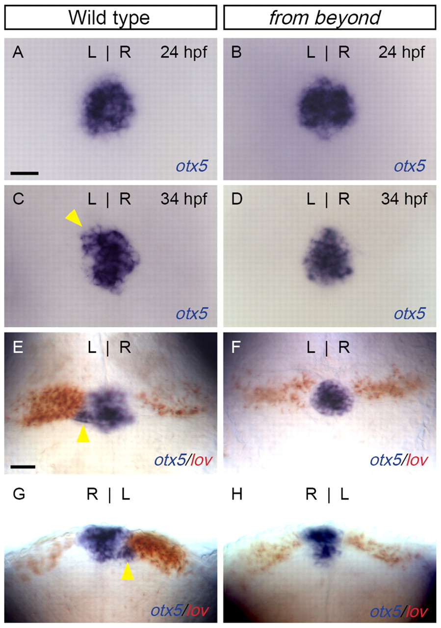
Fig. 3 Disruption of parapineal formation in fby mutant zebrafish larvae. (A-F) Dorsal and (G,H) frontal views of the epithalamus of (A,C,E,G) WT and (B,D,F,H) fby mutant larvae. (A,B) At 24 hpf, the pineal complex anlage labeled with otx5 (blue) appeared similar in WT embryos (A) and fby mutants (B). (C,D) However, by 34 hpf, when a parapineal was apparent in WT embryos (C), no parapineal organ developed in fby mutants (D). Arrowhead in C indicates the emerging parapineal organ. (E,G) By 4 dpf, WT larvae had a left-sided parapineal organ (arrowheads) and more cells expressed lov (red) in the left habenula. (F,H) In fby mutant larvae, no left-sided parapineal organ was apparent; rather, otx5-expressing cells were found below the pineal organ. The number of lov-expressing cells in the left habenula was reduced compared with WT. Scale bars: 25 μm.