
|
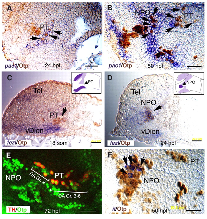
Fig. 2 Coordinated expression of pac1, fezl and Otp in the NPO and PT areas. (A-D,F) Sagittal paraffin sections (6 μm) through the NPO and PT (lateral view, anterior to the left). At the indicated stages of development, embryos were subjected to whole-mount in situ hybridization with probes directed against pac1 (A,B), fezl (C,D) or isotocin (it; F). Thereafter, specimens were sectioned and immunostained with an anti-Otp antibody. Schemes of fezl expression domain (dark purple) at the plane of the section are shown in C and D. Labeled RNA probes were detected in the cytoplasm, whereas Otp antibody exclusively labeled the nuclei. (E) Projected confocal Z-stack images of embryos (lateral view, anterior to the left) that were subjected to double immunofluorescence staining with monoclonal and polyclonal antibodies directed against tyrosine hydroxylase (TH) and Otp, respectively. The two prominent clusters of dopaminergic neurons, group 2 (Gr. 2) and groups 3-6 (Gr. 3-6), are indicated. hpf, hours post fertilization; NPO, neurosecretory preoptic area; PT, posterior tuberculum; vDien, ventral diencephalon. Scale bars: 20 μm in A-D; 40 μm in E,F.