
|
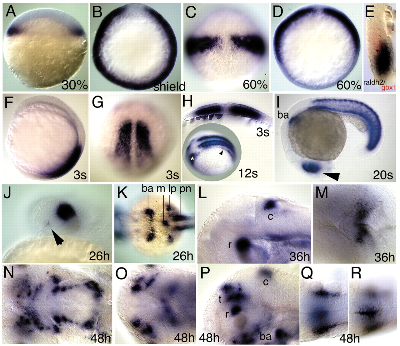
Fig. 2 Whole-mount in situ hybridisations showing raldh2 expression in the zebrafish embryo and larva at (A) 30% epiboly, (B-E) gastrula, during (F-I) somitogenesis, and (J-R) larval stages. (A) Marginal view, animal pole is upwards. (B,D) Animal view, dorsal is upwards. (C,G) Dorsal view, animal/anterior is upwards. (E) Sagittal section along the animal vegetal axis. (F) Lateral view, anterior is upwards. (H) Cross section perpendicular to anteroposterior axis. (H inset) Dorsolateral view, anterior is to the left. (I,J,L,P) Lateral view, anterior is to the left. (K,M,O,Q,R) Dorsal view, anterior is to the left. (N) Ventral view, anterior is to the left. Note (A) the continuous expression in the blastoderm margin, (B) the exclusion from the shield and (C,D,E) the restriction to the involuting paraxial mesendoderm at 60% epiboly. (E) The sagittal section also reveals positioning of gbx1 (K. L. and M. B., unpublished) (Rhinn and Brand, 2001) in the neuroectoderm adjacent to raldh2. (F,G,H,I) Expression in the somites during somitogenesis stages. (H inset) Expression in the lateral plate mesoderm (arrowhead) extends into the prospective caudal part of the branchial arch primordium (asterisk) at 12s. (I,J) Dorsal expression in the retina (arrowhead) at 20s (I) and 26 hours (J) and weak ventral expression near the choroid fissure at 26 hours (arrowhead). (K) Expression in the caudal part of the branchial arch primordium (ba); in a mesenchymal domain in the midline below the notochord (m), in the anterior part of the pronephric ducts (pn) and in the posterior fin bud- and lateral plate-mesoderm (lp) at 26 hours (arrowhead). (L,M) Expression in the retina (r) and cerebellum (c) at 36 hours. (N-P) Expression at 48 hours. (N) In patches indicating the developing arches; (O) in four domains in the tectum and (P) in the retina (r), cerebellum (c), tectum (t) and branchial arches (ba). (Q,R) Expression surrounding the neural tube at the level of somites 3 to 4.