
|
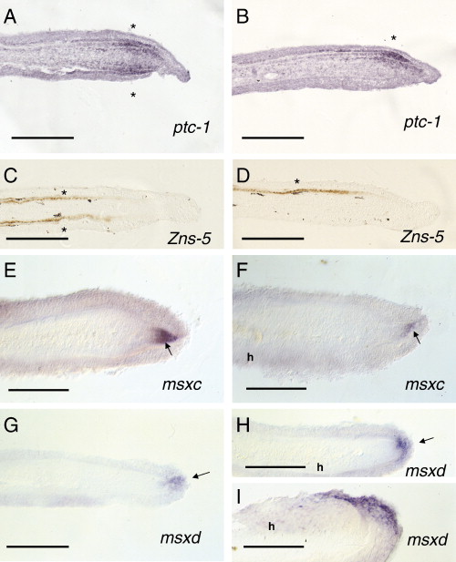
Fig. 3 In situ hybridization of fin blastema 4 or 5 days post-operation B. (A) ptc-1 expression is restricted to scleroblasts in both sides (asterisk) in control regenerating blastemas. (B) ptc-1 expression is observed in the scleroblasts regenerating the hemiray side (asterisk) in the experimental blastema. (C) Zns-5 expression is detected in both precursors of scleroblasts at both blastema sides (asterisks) during ray regeneration. (D) Zns-5 expression is exclusively detected in the regenerating hemiblastema at the level of both precursors of scleroblasts and scleroblasts (asterisk). (E, F) msxc expression is observed at a DMB (arrow in panel E) or DMH (arrow in panel F) in both control (E) and hemiray (h) blastemas. (G, H) msxd expression is restricted to the distalmost epidermis (arrow) covering the DMB (G) and DMH (H). (I) msxd expression is lateralized in the distal epidermis of a regenerating hemiblastema (H, I). h represents the side carrying a regenerating hemiray) Scale bar represents 100 μm (A–I).
Reprinted from Developmental Biology, 312(1), Murciano, C., Pérez-Claros, J., Smith, A., Avaron, F., Fernández, T.D., Durán, I., Ruiz-Sánchez, J., García, F., Becerra, J., Akimenko, M.A., and Marí-Beffa, M., Position dependence of hemiray morphogenesis during tail fin regeneration in Danio rerio, 272-283, Copyright (2007) with permission from Elsevier. Full text @ Dev. Biol.