
|
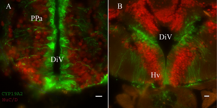
Fig. 7 Immunofluorescence staining with anti-CYP19A2 and anti-HuC/D. A: Detail from the anterior part of the parvocellular preoptic nucleus (PPa). CYP19A2 stains the cells lining the diencephalic ventricle (DiV), whereas anti-HuC/D stains the surrounding neurons. B: The anti-CYP19A2 antibody stains cell bodies in the area surrounding the ventral zone of the periventricular hypothalamus (Hv), and their processes that run down to the border of the brain that comes in contact with the pituitary (Pit). The cell bodies that are stained within the ventral zone of the periventricular hypothalamus (Hv) differ morphologically from the rest in the surrounding area as they are more elongated. Scale bar = 10 μm in A, 15 μm in B.