
|
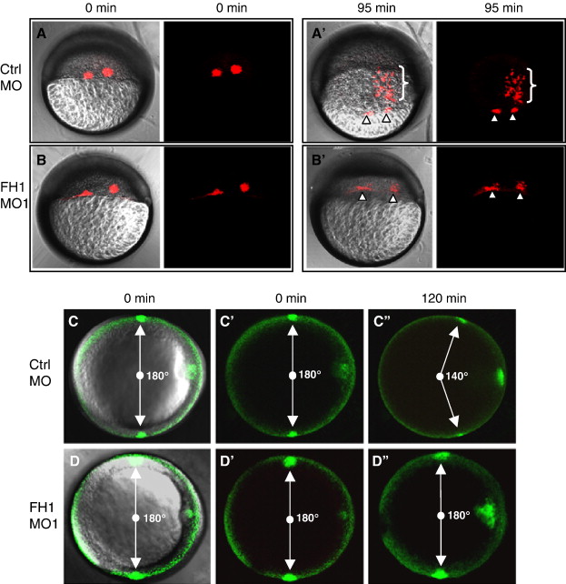
Fig. 3 FoxH1-depletion disrupts gastrulation movements. (A, B) FoxH1-depletion disrupts internalization of mesendoderm. Kaede protein was co-injected with 8 ng of the indicated MOs into wild type embryos. Cell labeling was performed by photoactivation of Kaede at 5 hpf (40% epiboly stage). Pictures were taken directly after labeling and 95 min later using lateral views. Primes (′) indicate different time points for the same embryos and bright field (left-hand panel) and fluorescent (right-hand panel) images are shown for each time point. Arrowheads point to the labeled cells remaining at the margin. Brackets in panel A2 indicate labeled cells in control embryos that internalize and migrate towards the animal pole. (C, D) FoxH1-depletion affects convergence.PA-GFP protein was co-injected with 8 ng of the indicated MOs into goosecoid-GFP embryos. Photolabeling of two groups of lateral cells was performed at shield stage using transgenic gsc-GFP as a reference point. Pictures were taken directly after labeling and 120 min later using animal pole views, with dorsal to the right. The angle between the labeled groups is indicated. Primes (2) indicate different time points for the same embryos and panels C and D are composite bright field/fluorescent views, whereas panels, C′, C″, D′ and D''' show the fluorescent channel alone. Abbreviations: FH1, FoxH1; ctrl, control.
Reprinted from Developmental Biology, 310(1), Pei, W., Noushmehr, H., Costa, J., Ouspenskaia, M.V., Elkahloun, A.G., and Feldman, B., An early requirement for maternal FoxH1 during zebrafish gastrulation, 10-22, Copyright (2007) with permission from Elsevier. Full text @ Dev. Biol.