
Image
|
Figure Caption
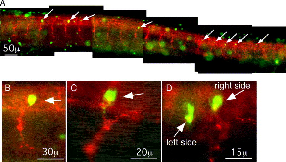
Fig. 5 Antibody staining for phosphorylated STAT3. (A) Mosaic image of a 27 h p.f. embryo stained with an antibody to phosphorylated STAT3 (green) and with ZNP1 (red) for primary motoneurons. White arrows mark neuronal cell bodies positive for phosphorylated STAT3. Green cells out of focus are EVL cells. (B–D) Higher magnification images of neuronal soma (white arrows) possessing phosphorylated STAT3. In all images, rostral is to the left, and dorsal is up.
Figure Data
Acknowledgments
This image is the copyrighted work of the attributed author or publisher, and
ZFIN has permission only to display this image to its users.
Additional permissions should be obtained from the applicable author or publisher of the image.
Reprinted from Developmental Biology, 296(1), Conway, G., STAT3-dependent pathfinding and control of axonal branching and target selection, 119-136, Copyright (2006) with permission from Elsevier. Full text @ Dev. Biol.