
|
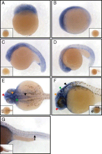
Fig. 3 Whole mount in situ analysis of lzic mRNA expression at various stages of early development. (A) lzic is ubiquitous at the 64-cell stage, lateral view animal pole to top. (B–D) lzic remains ubiquitous with increased anterior expression at 3 somites (B), 15 somites (C), and 24 somites (D), lateral views anterior to the left. (E–G) lzic expression at 24 h of development. (E) Dorsal view of zebrafish head shows strong brain expression, particularly in the telencephalon (red arrow), tectum (orange arrow), cerebellum (green arrow), and hindbrain (black arrow). (F) Lateral view (anterior to the left) showing the brain with arrows as in panel (E). (G) Lateral view of the tail, anterior to the left. Note expression in the notochord (black arrow). Insets show embryos stained with a sense lzic probe.
Reprinted from Developmental Biology, 283(2), Clements, W.K., and Kimelman, D., LZIC regulates neuronal survival during zebrafish development, 322-334, Copyright (2005) with permission from Elsevier. Full text @ Dev. Biol.