
|
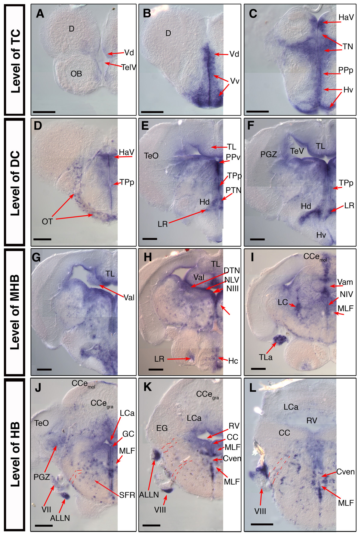
Fig. 6 nestin expression in the juvenile 28 dpf old zebrafish brain. Analysis of nestin expression in the brain of the 28 dpf zebrafish by whole mount in situ hybridization using nestin anti sense mRNA probes on whole dissected brain. (A-L): selected 50 μm cross sections from a serially sectioned brain, the rostrocaudal levels of the sections are indicated at left; dorsal up. Pictures show only the left half of the brain. Dashed lines in J, K, L indicate the tracts of the cranial nerves. Abbreviations: ALLN: anterior lateral line nerve; CCe: corpus cerebelli; Cven: commissura ventralis rhombencephali; D: dorsal telencephalic area; DiV: diencephalic ventricle; EG: eminentia granularis; GC: griseum centrale; HaV: ventral habenular nucleus; Hc, Hd, Hv: caudal, dorsal, ventral zone of the periventricular hypothalamus; LC: locus coeruleus; LCa: locus caudalis cerebelli; LR: lateral recess of the DiV; MLF: medial longitudinal fascicle; NIII: oculomotor nucleus; NIV: trochlear nucleus; NMLF: nucleus of MLF; NLV: nucleus lateralis valvulae; OT: optic tract; PGZ: periventricular gray zone of the optic tectum; PPa, PPp: parvocellular preoptic nucleus, anterior, posterior part; PPv: periventricular pretectal nucleus, ventral part; PTN: posterior tuberal nucleus; RV: rhombencephalic ventricle; TeO: optic tectum; TeV: tectal ventricle; TelV: telencephalic ventricle; TL: Torus longitudinalis; TLa: torus lateralis; TN: thalamic nuclei; Val, Vam: lateral, medial division of valvula cerebelli; Vd, Vv: dorsal, ventral nucleus of ventral telencephalic area; TPp: periventricular nucleus of the posterior tuberculum; III: oculomotor nerve; VII: facial nerve; VIII: octaval nerve; Scale bars: 100 μm.