
|
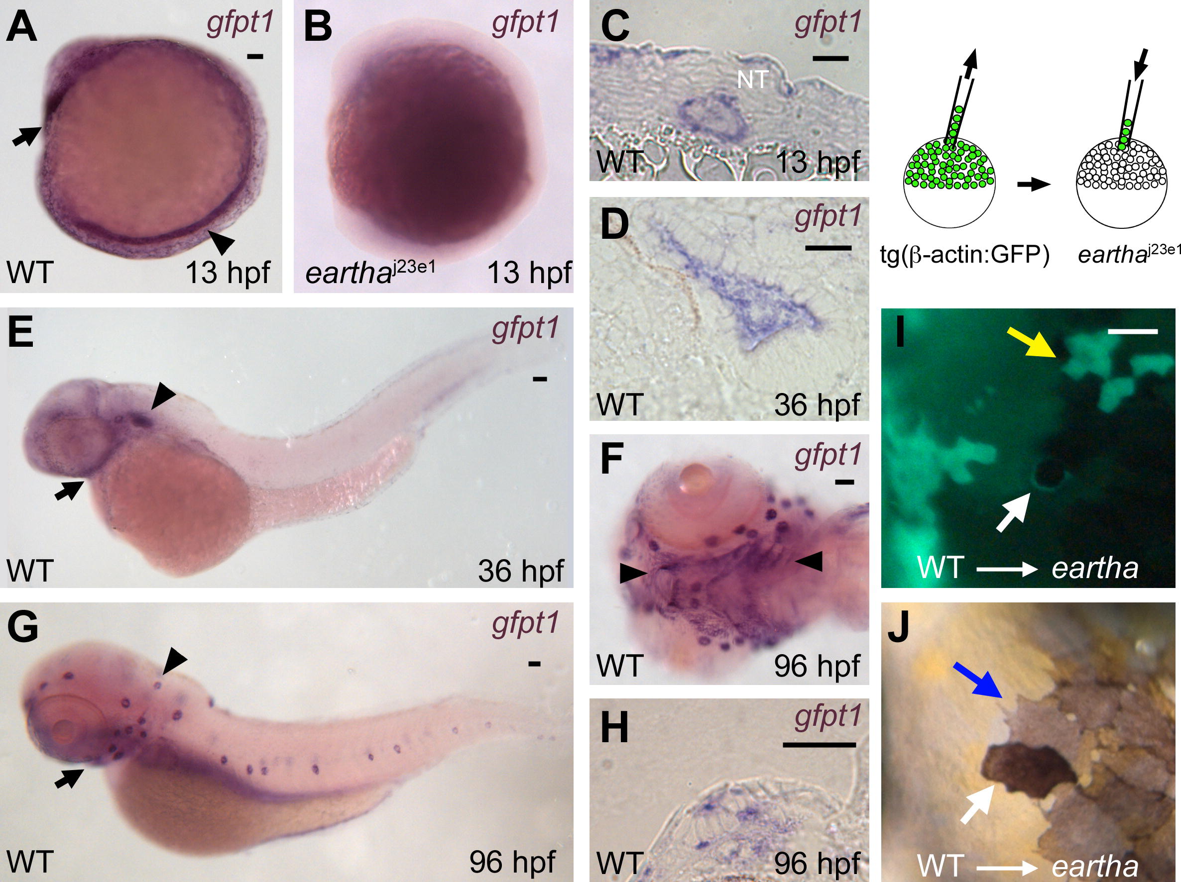
Fig. 5 gfpt1 Is Expressed in Notochord, Ear, and Pharyngeal Arches, and Acts Cell Autonomously in Melanocytes to Promote Melanocyte Darkening (A–H) ISH reveals high expression of gfpt1 in the notochord (arrowhead in [A]) and the pillow (polster, arrow in [A]) at 13 hpf. No or very few gfpt1 transcripts were detected in the earthaj23e1 mutants at 13 hpf (B). A cross section (C) shows the gfpt1 expression in notochord at 13 hpf. gfpt1 is expressed in the ear (arrowhead in [E]) and pharyngeal arches (arrow in [E]) at 36 hpf. gfpt1 expression in the sensory epithelium of the ear is shown in a cross section (D). At 96 hpf, gfpt1 is expressed in the lateral line neuromasts (arrowhead in [G]) and the pharyngeal arches (arrow in [G] and arrowheads in [F]). A cross section (H) shows the gfpt1 expression in the neuromasts. (I–J) Chimeric animals were generated by transplanting wild-type tg(β-actin:GPF) donor cells into earthaj23e1 embryos at midblastula stage. A donor (GFP+) melanocyte in the eartha larvae is revealed following incubation in 0.1% epinephrine to induce melanosome contraction (white arrow in [I]). In the uncontracted state, the GFP+ (donor-derived) melanocyte appears darkly pigmented, in contrast to the lightly pigmented neighboring melanocytes (GFP-) that are derived from host (blue arrow in [J]). Yellow arrow in (I) indicates donor-derived skin cells (GFP+) that are not associated with darkened melanocytes. Scale bars: in (A), 50 μm for (A and B); in (C and D) and (H), 20 μm; in (E–G), 50 μm; in (I), 50 μm for (I and J).