
|
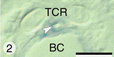
Fig. 2 dlx gene expression during cranial bone development in zebrafish larvae in Figures 2-23. Transverse, 6-μm-thick sections of whole-mount hybridized specimens (Figs. 2, 3, 2, 8, 9, 10, 14, 15, 16, 20, 21) with their respective 2-μm-thick toluidine blue-stained reference section (5, 6, 7, 11, 12, 13, 17, 18, 19, 22, 23). Legends to all reference sections are identical to those of the hybridized sections. BC, buccal cavity; CB4, ceratobranchial cartilage 4; CB5, ceratobranchial cartilage 5; CH, ceratohyal cartilage; E, eye; HM, hyomandibular cartilage; MC, Meckel's cartilage; O, opercular bone; PQ, palatoquadrate cartilage; TC, trabecula communis; TCR, trabecula cranii. Scale bars = 20 μm.At 3 days postfertilization (dPF). Expression of dlx3b in the osteoblasts surrounding the parasphenoid bone (arrowhead).