
Image
|
Figure Caption
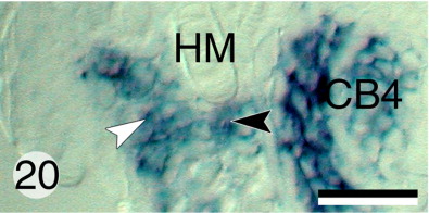
Fig. 20 At 3 dPF. Expression of dlx5a in the mesenchymal cells (black arrowhead) ventral to the hyomandibular cartilage where the perichondral hyomandibular bone will form. Lateral to the hyomandibula, dlx5a expression is strong in the osteoblasts surrounding the opercular bone (white arrowhead). Strong expression is also observed in mesenchymal cells of the fourth branchial arch. Probably this specimen is slightly advanced, given the observation that this expression connected to the hyomandibular usually only starts at 4 dPF.
Figure Data
Acknowledgments
This image is the copyrighted work of the attributed author or publisher, and
ZFIN has permission only to display this image to its users.
Additional permissions should be obtained from the applicable author or publisher of the image.
Full text @ Dev. Dyn.