
|
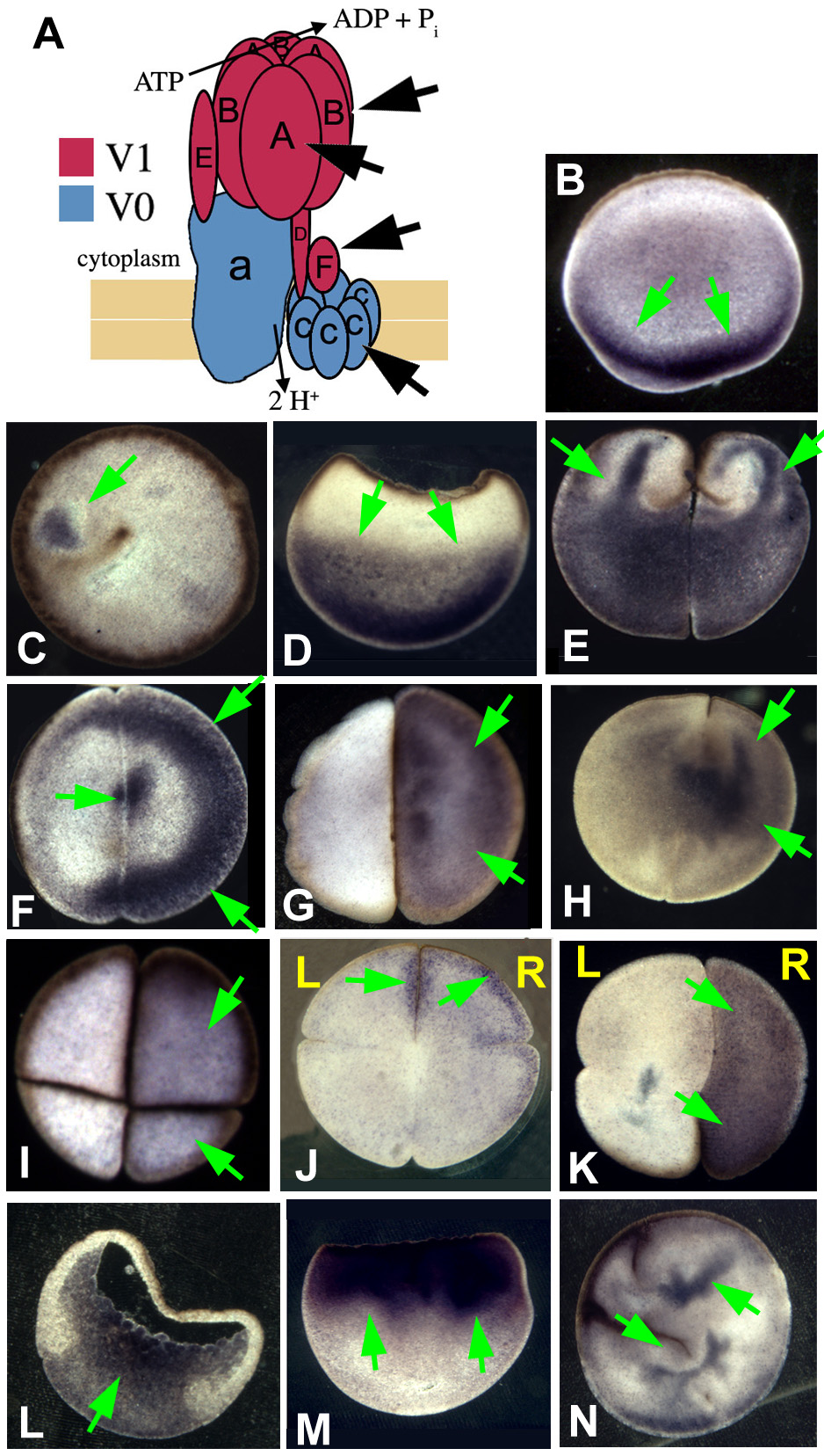
Fig. S1 Immunohistochemistry for H+-V-ATPase subunits in early Xenopus embryos. Cartoon showing the arrangement of subunits in the H+-V-ATPase (after Nishi 2002). (B) Subunit A in an AV section (parallel to the animal-vegetal axis) of an unfertilized egg; subunits are found localized in the vegetal cortex. (C) A just-fertilized egg stained with anti-subunit F. A small area of staining can be seen next to the brown curve of pigment granules marking the path of sperm entry. (D) In post-rotation zygotes, staining for subunit A has moved up into the vegetal cytoplasm (AV section). (E-H) Two-celled embryos stained for subunits F and A. E illustrates a commonly seen pattern: subunit F staining appears to form fingers that reach up from the vegetal to the animal cytoplasm. In flat sections (perpendicular to the AV axis), staining is usually asymmetric and heavy near the cell membrane: (F) subunit F; (G) subunit A; (H) subunit A revealed with a different primary antibody. (I) Four-cell embryos, flat sections: (I) subunit A; (J) oriented section, subunit B. Staining is on the right hand side. (K) Flat section, oriented eight-cell embryo stained for subunit B. Staining is on the right hand side. (L) AV section through gastrula-stage embryo stained for subunit A. By this stage, staining is mostly uniformly distributed with respect to the LR axis. (M) Embryo treated with cytochalasin B to disrupt actin filaments, fixed when controls reached the two cell stage (no cleavage furrows form in actin-disrupted embryos). Staining for subunit B shows that the normal vegetal-plus-fingers localization of H+-V-ATPase subunits (compare to E) has become entirely animal. (N) Embryo treated with latrunculin to disrupt actin filaments, imaged when controls reached the four-cell stage. Subunit A staining reveals that localization has been completely disrupted (compare with I or J). Although the patterns presented in this figure match those found with the anti-subunit A (Fig. 3), the antibodies used for this figure, although highly specific in mammals, did not, in our hands, work for westerns.