
|
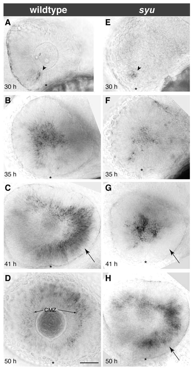
Fig. 6 Delay of the ath5 wave caused by loss of shh signaling. Timecourse of ath5 expression in wild-type or mutant embryos derived from a syu t4/+ intercross. (A-D) In wild type, the wave front is: in the ventronasal retina at 30 hpf (A); in the central retina at 35 hpf (B); and in the temporal retina at 41 hpf (C). At 50 hpf (D), the wave is over and ath5 expression is seen only in the secondary retinal growth zone, the ciliary marginal zone (arrows in D). (E-H) In syu mutants, wave initiation is normal (E) but subsequently becomes delayed. About half of mutants show normal spread of ath5 wave to central retina by 35 hpf (F); the other half fail to show expression in central retina by 35 hpf (not pictured; see Fig. 7B). By 41 hpf (G) the delay phenotype is fully penetrant – note absence of ath5 expression in temporal retina at 41 hpf (arrow in G). By 50 hpf (H), ath5 expression reaches the temporal retina and has cleared from the central retina, much like in wild type at 41 hpf (arrows in H,C). Nasal/anterior is left and dorsal is up in all panels. Asterisks mark the choroid fissure. Scale bar: 50 µm. CMZ, ciliary marginal zone.