
Fig. 2
- ID
- ZDB-FIG-240111-10
- Publication
- Tan et al., 2023 - Fgf, Hh, and pax2a differentially regulate expression of pax5 and pou3f3b in vestibular and auditory maculae in the zebrafish otic vesicle
- Other Figures
- All Figure Page
- Back to All Figure Page
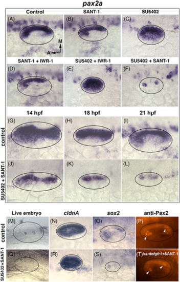
Critical roles for Fgf and Hh in regulating pax2a and sensory development. (A-T) Dorsolateral views of the otic placode or vesicle at various stages. (A-F) Images showing pax2a expression at 24 hpf following treatment at 12 hpf with inhibitors for Hh, Wnt, and Fgf (listed at top). IWR-1 (10 μM) was used to block Wnt. (G-L) Time course of pax2a expression in wild-type (DMSO treated) or SU5402 + SANT-1 treated embryos. Drugs were administered at 12 hpf and embryos were fixed at the timepoints listed at top. (M, Q) Images of live embryos at 24 hpf following treatment with DMSO (control) or SU5402 + SANT-1. (N, R) Expression of cldnA at 24 hpf in control and drug treated embryos. (O, S) Expression of sox2 at 24 hpf in control and drug treated embryos. (P, T) Anti-Pax2 staining at 30 hpf after blocking Fgf and Hh. Here Fgf was blocked by activating hs:dnfgfr1 to avoid intense background fluorescence caused by SU5402. Embryos were heat shocked at 12 hpf for 1 h at 39°C followed by immediate addition of SANT-1 (150 μM). Arrowheads mark elevated staining in tether cell nuclei. |