FIGURE

Fig. 1

ID
ZDB-FIG-231208-27
Publication
Celeghin et al., 2023 - A novel DSP zebrafish model reveals training- and drug-induced modulation of arrhythmogenic cardiomyopathy phenotypes
Other Figures
All Figure Page
Back to All Figure Page
Fig. 1

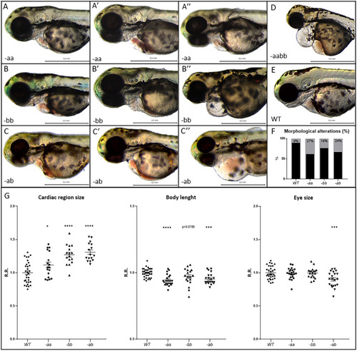
Cardiac alterations and developmental delay in Dsp mutant lines.

AF Mutant embryos (AC″, D) display cardiac alterations, compared to WT (E). Homozygous -aa (AA″) and -bb (BB″) mutant hearts appear dilated and/or structurally altered, with cardiac pericardial effusion and/or hemopericardium in the cardiac region. Double heterozygotes -ab (CC″) show a more serious phenotype than -aa and -bb lines. Double homozygotes -aabb display the most severe phenotype (D). F Percentage of heart alteration in different genotypes. Sample size: n = 100. G Cardiac size analysis shows that all mutants present heart dilation. Body length and eye size measurements indicate developmental delay in mutants, especially in the -ab condition. All embryos are at 3 dpf in lateral view, anterior to the left. Sample size Cardiac region size: WT n = 31; -aa n = 18; -bb n = 16; -ab n = 16. Sample size Body length: WT n = 32; -aa n = 22; -bb n = 19; -ab n = 21. Sample size Eye size: WT n = 32; -aa n = 22; -bb n = 19; -ab n = 21. R.R. relative ratio. Error bars: SEM. *p < 0.05; **p < 0.01; ***p < 0.001; ****p < 0.0001. Test: One-way ANOVA followed by Tukey’s test.

Expression Data

Expression Detail
Antibody Labeling
Phenotype Data

Phenotype Detail
Acknowledgments
This image is the copyrighted work of the attributed author or publisher, and ZFIN has permission only to display this image to its users. Additional permissions should be obtained from the applicable author or publisher of the image. Full text @ Cell Death Discov