
Fig. 4
- ID
- ZDB-FIG-231128-25
- Publication
- Pownall et al., 2023 - Chromatin expansion microscopy reveals nanoscale organization of transcription and chromatin
- Other Figures
- All Figure Page
- Back to All Figure Page
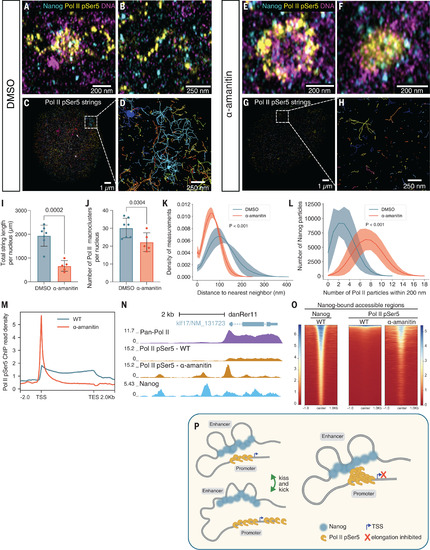
Nanog-bound enhancers and Pol II-bound promoters are kicked apart by transcription elongation. (A and B) Representative images of Nanog and Pol II pSer5 in dimethyl sulfoxide (DMSO)–treated embryos at 4 hpf. n = 6 nuclei from 2 embryos. (C and D) Visualization of Pol II pSer5 strings identified in DMSO-treated embryos at 4 hpf. (E and F) Representative images of Nanog and Pol II pSer5 in α-amanitin–treated embryos. (G and H) Visualization of Pol II pSer5 strings identified in α-amanitin–treated embryos. (I) Quantification of total Pol II pSer5 string length per nucleus in DMSO- and α-amanitin–treated embryos. P = 0.0002; unpaired t test. (J) Quantification of the number of Pol II pSer5 macroclusters detected in DMSO- and α-amanitin–treated embryos. P = 0.0304; unpaired t test. (K) Density plot of the distance to nearest neighbor for Nanog to Pol II pSer5 particles in DMSO- and α-amanitin–treated embryos. n = 221,335 and 229,706 distances, respectively. P < 0.001; Mann-Whitney U test. (L) Histogram showing the number of Pol II pSer5 particles within 200 nm of each Nanog particle in DMSO- and α-amanitin–treated embryos. P < 0.001; Mann-Whitney U test. (M) Line plot showing Pol II pSer5 binding across gene bodies ±2 kb at zygotic genes in wild-type (WT) and α-amanitin–treated embryos. TES, transcription end site TSS, transcription start site. (N) Representative genome tracks of pan-Pol II, Pol II pSer5, and Nanog binding showing accumulation of Pol II pSer5 at the promoter and Nanog-bound enhancers in the presence of α-amanitin. (O) Heatmaps showing Pol II pSer5 binding at Nanog-bound accessible regions in WT and α-amanitin–treated embryos. Regions are ranked by Nanog ChIP-seq signal. (P) Schematic showing the kiss-and-kick model. |