FIGURE

Fig 3

ID
ZDB-FIG-231118-3
Publication
Veen et al., 2023 - Her6 and Prox1a are novel regulators of photoreceptor regeneration in the zebrafish retina
Other Figures
All Figure Page
Back to All Figure Page
Fig 3

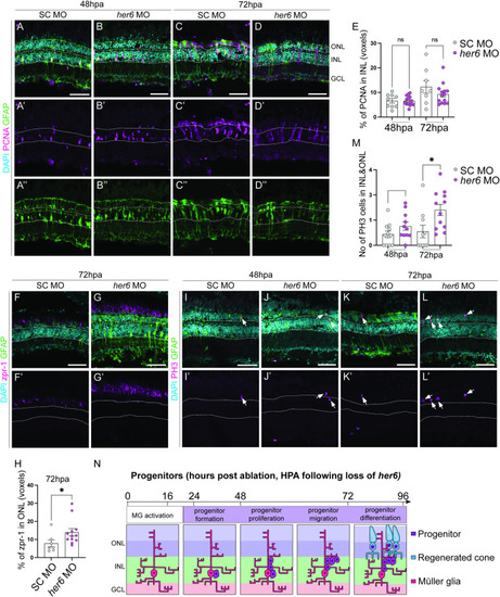
Loss of Her6 increases PR production at 72 hours post ablation.

(A-D”) In adult retina, at 48 and 72 hours post ablation (hpa), the number of proliferating cells marked by PCNA (pink) and Müller glia (MG) cells marked by glial fibrillary acidic protein transgene expressing (GFAP, green) are not significantly altered between standard control (SC, 48hpa n = 9, 6.621 ±0.9034, 72hpa n = 8, m = 12.27 ±2.579) and her6 morpholino (MO, 48 hpa n = 14, m = 6.59 ±0.5219, 72 hpa n = 14, m = 9.299 ±1.1) electroporated samples quantified in (E). (F-G’) In her6 MO samples, there was a significantly increase in zpr1 (pink, n = 12, 13.97 ±2.202) labelled cone photoreceptors compared to SC (n = 7, 7.809 ±1.938), quantified in H. (I-L) There was also an increase in phospho-histone 3 (PH3, pink) marking mitotic cells in the her6 MO (48hpa, n = 11, 1.519 ±0.3166, 72hpa n = 12, 2.806 ±0.4442) compared to SC (48hpa n = 11, 0.8812 ±0.2671, 72hpa (SC n = 10, 1.102 ±0.4956) quantified in M. In all micrographs DAPI is used to label nuclei cyan. Data are represented as mean ± SEM. *p < 0.05. Scale bars: 50 μm. (N) Summary of regenerative phases and timeline in her6 MO electroporated fish showing an increase in mature photoreceptor regeneration.

Expression Data

Expression Detail
Antibody Labeling
Phenotype Data

Phenotype Detail
Acknowledgments
This image is the copyrighted work of the attributed author or publisher, and ZFIN has permission only to display this image to its users. Additional permissions should be obtained from the applicable author or publisher of the image. Full text @ PLoS Genet.