
Fig. 2
- ID
- ZDB-FIG-231106-65
- Publication
- Mytlis et al., 2023 - The many faces of the bouquet centrosome MTOC in meiosis and germ cell development
- Other Figures
- All Figure Page
- Back to All Figure Page
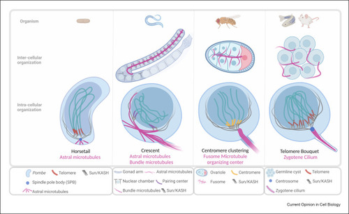
Anchoring strategies of the bouquet MTOC machinery. Bouquet and bouquet-analogous configurations are shown in different species. In all cases, telomeres (S. pombe, zebrafish, and mouse), centromeres (Drosophila), or pairing centers (C. elegans) are bound to Sun/KASH complexes on the NE and moved by dynein on perinuclear MTs as shown in Figure 1. The potential anchor (magenta) and its intercellular and intracellular organizations are shown for each species. In S. pombe, the centrosome (spindle pole body, SPB) is anchored through MT to the cytoplasmic membrane. In the C. elegans gonads, germ cells are organized in nuclear chambers (see text), and perinuclear MTs are anchored to the cytoplasmic membrane of nuclear chambers. Some perinuclear MTs extend into the rachis and join a common network of bundles that runs through the gonadal arm, the anchoring functions of which have yet to be tested. In Drosophila, perinuclear MTs are prominently organized from the fusome, which runs through the germline cyst in the egg chamber and could function as an anchoring scaffold. In zebrafish, the bouquet centrosome is anchored by the zygotene cilium, which is likely in mice as well. |