|
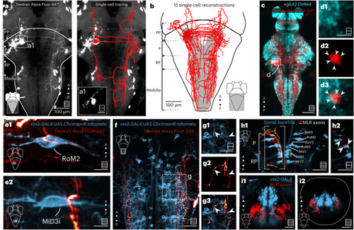
MLR neurons project bilaterally to V2a RSNs forming putative axosomatic synapses in the P and RP areas and axodendritic synapses in the medulla. a,a1, Left: forsal view of a Z-stack showing the projection pattern of an MLR neuron that was electroporated with Dextran Alexa Fluor 647. Right: reconstruction of the arborization pattern. b, Reconstruction of electroporated neurons with somata located in the MLR locus (7 fish, 15 neurons). c, The MLR contains many glutamatergic neurons projecting to the reticular formation. Scale bar, 100 µm. d1–d3, The single plane revealed that many MLR electroporated neurons (red) were glutamatergic, as shown by the overlap with the Tg(vglut2:DsRed) transgenic line (cyan) (7 fish, 14 neurons). Scale bar, 20 µm. e, Electroporation of MLR neurons (red) in Tg(vsx2:GAL4;UAS:ChrimsonR-tdtomato) (blue) transgenic fish. Putative connections of the MLR to an ipsilateral P RSN (e1) and a contralateral RP RSN (e2). Scale bars, 10 µm. f,g, Z-stack of the medulla region (f) showing the typical projection pattern on the MLR neurons to the lateral dendritic area (g1–g3). h1, Dorsal view of a Z-stack of spinal backfills labeling (blue) and two examples of MLR neurons (orange and red) taken from the single-cell atlas mapzebrain. h2, Single plane of the region denoted in h1 showing the axons of the MLR neurons reaching the soma of the P and RP RSNs. i, Coronal view of a Z-stack of V2a neurons (blue) and all MLR neurons recovered from the single-cell atlas mapzebrain (red) in the RP region (i1) and caudal medulla (i2). Scale bars, 40 µm. In all panels, maximum projection Z-stacks are schematized in the bottom right corner, with squares including multiple lines and single optical sections schematized with squares including a single line. Source data
|