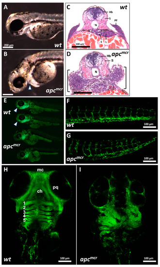
apc is required for the CNC contribution to craniofacial structures: (A) Wild-type (WT) and (B) apcmcr/mcr larvae at ~30 somites (50 hpf). Arrowhead indicates the accumulation of amorphous tissue anterior to the heart and yolk mass. Scale bars in (A,B) are 200 µm. (C,D) Hematoxylin-eosin staining of WT (C) and apcmcr/mcr (D) larvae sectioned at the level of the hindbrain. Hb: hindbrain; S: somite; N: notochord; E: esophagus; L: liver; PF: pectoral fin. Scale bars in (C,D) are 100 µm. (E) Fli1-GFP epifluorescence of WT (top two larvae) and apcmcr/mcr larvae (lower two larvae; 30-somite stage). Arrowheads for WT larvae indicate anterior extent of GFP+ cells in the head, which is reduced or absent in mutants. (F,G) Confocal image of WT; Fli1-GFP (F) and apcmcr/mcr; Fli1-GFP (G) larvae at 60 hpf, showing the vascular endothelium of trunk. (H,I) Confocal image of Fli1-GFP in WT (H) and apcmcr/mcr (I) larvae at 60 hpf, showing CNC and vascular endothelium in branchial arches and head. mc: Meckel’s cartilage; pq: palatoquadrate cartilage; ch: ceratohyal cartilage. Numbers indicate gill arches 1–5.
|
