
Fig. 1
- ID
- ZDB-FIG-230522-24
- Publication
- Emmerich et al., 2023 - Nanoparticle-based targeting of microglia improves the neural regeneration enhancing effects of immunosuppression in the zebrafish retina
- Other Figures
- All Figure Page
- Back to All Figure Page
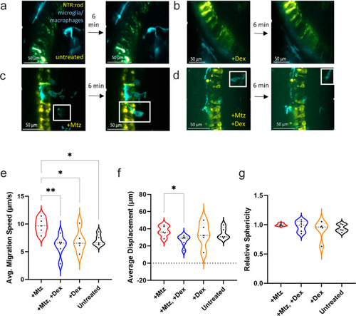
Microglia reactivity is altered in response to post-ablation Dex treatment.
a–d Image stills 6 min apart from AO-LLSM imaging of 5 or 6 dpf transgenic lines labeling NTR-expressing rods (yellow) and microglia/macrophages (cyan). Four conditions were imaged: non-ablated “untreated” control (A), non-ablated, “+Dex” (B), rod cell ablated, “+Mtz” (C), and rod cells ablated with Dex treatment, “+Mtz, +Dex” (D). In all images, the inner nuclear layer is to the right of the NTR-YFP rod cells. e–g Imaris quantification of average migration speed (μm/second), average displacement (μm), and relative sphericity of microglia in each larva, sample sizes for each plot from left to right: 5, 6, 5, 6 with a total of 66 microglia analyzed. See Supplementary Movies 1-4 corresponding to stills A-D, respectively. Asterisks indicate statistically significant differences between the indicated groups (*p ≤ 0.05, **p ≤ 0.01), all other comparisons were not statistically significant. Lines within the violin data for each condition for all plots indicate lower quartile (bottom), median (middle line), and upper quartile (top). |