
FIGURE
Fig. 5
- ID
- ZDB-FIG-230327-40
- Publication
- Shin et al., 2022 - Three-dimensional fluorescence microscopy through virtual refocusing using a recursive light propagation network
- Other Figures
- All Figure Page
- Back to All Figure Page
Fig. 5
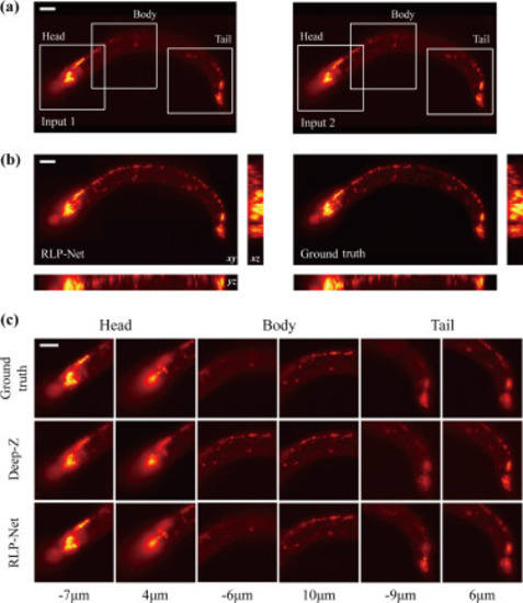
Fig. 5. Virtual refocusing of C. elegans images. (a) Two adjacent images used as the input. Each region-of-interest shows head ganglia, body, and tail ganglia, respectively. (b) MIPs of the reconstructed volume using RLP-Net (left) and the ground-truth (right). In each panel, lateral () MIP (top-left), axial () MIP (bottom-left), and axial () MIP (right) are shown. (c) The ground truth images (1st row) are compared with the refocusing results from Deep-Z (2nd row) and RLP-Net (3rd row). Different neurons are in focus at different axial locations. Scale bars, 20 m. |
Expression Data
Expression Detail
Antibody Labeling
Phenotype Data
Phenotype Detail
Acknowledgments
This image is the copyrighted work of the attributed author or publisher, and
ZFIN has permission only to display this image to its users.
Additional permissions should be obtained from the applicable author or publisher of the image.
Reprinted from Medical image analysis, 82, Shin, C., Ryu, H., Cho, E.S., Han, S., Lee, K.H., Kim, C.H., Yoon, Y.G., Three-dimensional fluorescence microscopy through virtual refocusing using a recursive light propagation network, 102600, Copyright (2022) with permission from Elsevier. Full text @ Med. Image Anal.