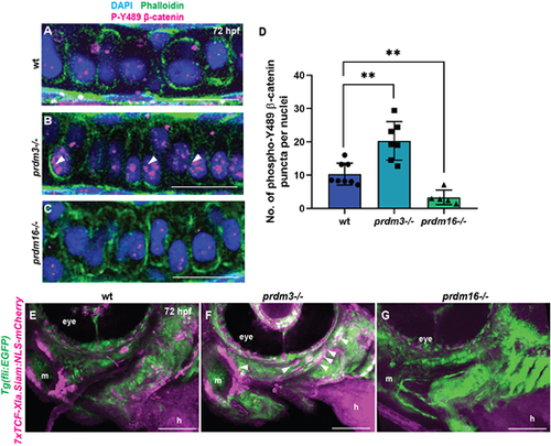
Prdm3 and Prdm16 control β-catenin stabilization and localization in craniofacial chondrocytes. (A-D) Wild-type (wt), prdm3−/− and prdm16−/− zebrafish embryos were collected at 75 hpf and immunostained for nuclear β-catenin (phosphorylated tyrosine 489) and counterstained with phalloidin and DAPI (A-C). Shown are high-magnification lateral images of the palatoquadrate. Increased nuclear β-catenin (magenta) was observed in prdm3−/− (white arrowheads in B), which was significantly reduced in prdm16−/− (C). (D) Quantification of the number of β-catenin puncta across ten nuclei per individual and averaged across at least five embryos per genotype; mean±s.d. Scale bars: 50 µm. (E-G) prdm3−/− and prdm16−/− mutant lines were crossed into the Wnt reporter line Tg(7xTCF-Xla.Siam:NLS-mCherry) to assess Wnt-responsive cells. Shown are representative lateral-ventral views of 75 hpf wild-type (E), prdm3−/− (F) and prdm16−/− (G) embryos. Increased Wnt-responsive cells were identified in the pharyngeal arch tissues of prdm3−/− (F) (white arrowheads), with a dramatic decrease in Wnt-responsive cells in prdm16−/− (G) mutants compared with wild type (E). h, heart; m, mouth. Scale bars: 100 µm. **P≤0.005 (unpaired, two-tailed Student's t-test).
|
