
Fig. 3
- ID
- ZDB-FIG-230308-5
- Publication
- Subkhankulova et al., 2023 - Zebrafish pigment cells develop directly from persistent highly multipotent progenitors
- Other Figures
- All Figure Page
- Back to All Figure Page
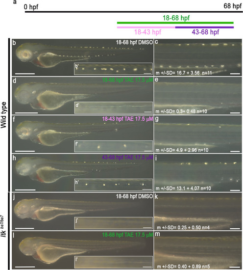
Schematic showing time window treatment (a). Embryos were treated with DMSO (b and c) or 17.5 μM TAE-684 (d–i) during defined time periods: 18-68 hpf (‘control’, b-c), 18-68 hpf (‘full’, d-e), 18-43 hpf (‘early’, f-g) and 43-68hpf (‘late’, h-i). Iridophores in the dorsal stripe were detected through their endogenous reflectivity; PTU-treatment from 24 hpf was used to suppress melanisation. Note how ‘early’ inhibition of Ltk reduces the number of iridophores (specification defect), whilst ‘late’ inhibition prevents the enlargement of iridophores (proliferation or differentiation). Homozygous ltk mutants (ltkba7/ba7) treated with DMSO (j and k) or 17.5 μM TAE-684 (l-m) during 18-68 hpf were indistinguishable, showing no more than occasional iridophores in the dorsal stripes, and similar to WT embryos treated in the same time window (d and e). All images were taken left side view with dorsal to the top. b–hn = 10 embryos on each condition; n = 3 independent experiments. jn = 4 embryos, ln = 5 embryos; n = 3 independent experiments. Source data are provided as a Source Data file. |