
Fig. 3
- ID
- ZDB-FIG-230213-3
- Publication
- Chiang et al., 2023 - HyU: Hybrid Unmixing for longitudinal in vivo imaging of low signal-to-noise fluorescence
- Other Figures
- All Figure Page
- Back to All Figure Page
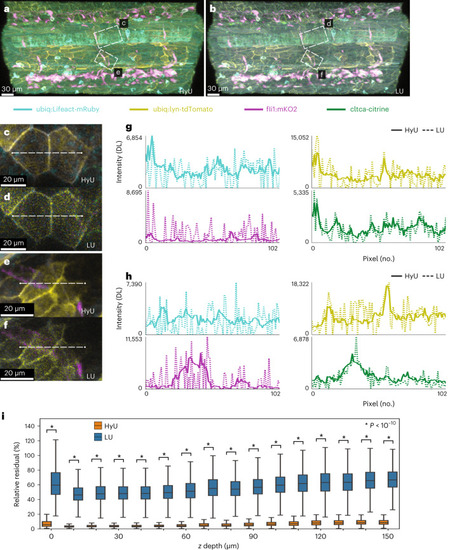
HyU enhances unmixing for low-signal in vivo multiplexing and achieves deeper volumetric imaging.
a,b, HyU volumetric renderings (a) compared to those of LU (b) for the trunk portion in a four-color zebrafish. The four labels in the fish are Gt(cltca-citrine);Tg(ubiq:lyn-tdTomato;ubiq:Lifeact-mRuby;fli1:mKO2), respectively labeling clathrin-coated pits (green), membrane (yellow), actin (cyan) and endothelial (magenta). c–f, Zoom-in views of insets from a and b. Scale bar, 20 µm. g,h, Intensity line plots of each of the four results signals for HyU (solid) and LU (dashed) demonstrate the improved profiles with greatly reduced noise peaks in HyU as compared to LU. Intensities are scaled by the maximum of each unmixed channel. DL, digital level. i, Box plots of the relative residual values as a function of z depth for HyU and LU highlight the improvements in the unmixing results. HyU has an unmixing residual of 6.6 ± 5.3% compared to that of LU at 58 ± 17%. The average amount of residual is ninefold lower in HyU with narrower variance of residual. An independent two-sample t-test was used with n = 5.2 × 105 pixels for each z slice. Center shows median; box shows first and third quartile; whiskers show 1.5× first and third quartiles; min/max are not shown. The sample depicted is representative of 28 experimental sessions each with three to five biological replicates, yielding similar results. |