
Fig. 2
- ID
- ZDB-FIG-230204-29
- Publication
- Brown et al., 2021 - A novel gene trap line for visualization and manipulation of erbb3b+ neural crest and glial cells in zebrafish
- Other Figures
- All Figure Page
- Back to All Figure Page
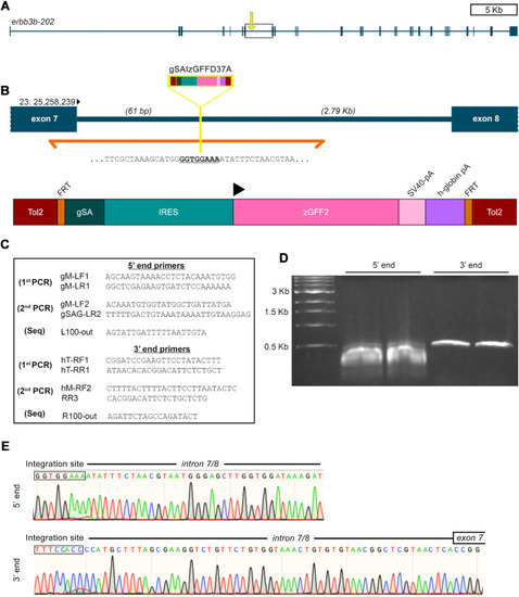
Fig. 2. gSAIzGFFD37A is located in erbb3b intron 7/8. (A) erbb3b transcript structure with exons shaded as rectangles. (B) Zoomed in view of intron 7/8 from box and arrow in panel (A). Inverse PCR sequencing (orange) revealed that the insertion site (yellow) is 61 bp downstream of erbb3b exon 7 and 2.9 kb upstream of exon 8 on the + strand of chromosome 23 (Tol2 = Tol2 element; FRT = FRT site; gSA = splice acceptor; IRES = internal ribosome entry sequence; zGFF2 = Gal4 transcription activator; SV40-pA = SV40 polyadenylation signal sequence; h-globin pA = globin polyadenylation signal sequence). (C) Primers used for inverse PCR and sequencing. (D) Gel electrophoresis of products from the second round of inverse PCR. (E) Chromatogram of initial sequence reads from purified 5′ end bands (top) and 3′ end bands (bottom) starting from the gSAIzGFFD insertion site. |
Reprinted from Developmental Biology, 482, Brown, E.A., Kawakami, K., Kucenas, S., A novel gene trap line for visualization and manipulation of erbb3b+ neural crest and glial cells in zebrafish, 114-123, Copyright (2021) with permission from Elsevier. Full text @ Dev. Biol.