FIGURE

Fig. 2

ID
ZDB-FIG-221209-21
Publication
Moss et al., 2021 - Autophagy coordinates chondrocyte development and early joint formation in zebrafish
Other Figures
All Figure Page
Back to All Figure Page
Fig. 2

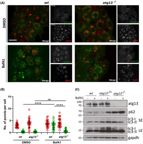
atg13 mutants show reduced autophagy flux. (A) Representative single confocal z‐slices of epidermal cells taken from LysoTracker stained Tg(cmv:gfp‐map1lc3;atg13) wt and mutant larvae, at 4 dpf following treatment with DMSO or 100 nM Bafilomycin for 3 h. Scale bars = 10 µm. (B) Quantification of number of lysosomal (red) and GFP‐Lc3 (green) puncta per cell. Two‐way ANOVA performed for each; ****p < .0001. (C) Representative immunoblot of atg13 wt, heterozygous and mutant larvae at 5 dpf following treatment with DMSO or 100 nM Bafilomycin for 3 h. LE, long exposure; SE, short exposure. Molecular weight markers indicated on right hand side of immunoblots

Expression Data
Antibodies:
Fish:
Condition:
Anatomical Terms:
Stage Range: Day 4 to Day 5

Expression Detail
Antibody Labeling
Phenotype Data
Fish:
Condition:
Observed In:
Stage Range: Day 4 to Day 5

Phenotype Detail
Acknowledgments
This image is the copyrighted work of the attributed author or publisher, and ZFIN has permission only to display this image to its users. Additional permissions should be obtained from the applicable author or publisher of the image. Full text @ FASEB J.