Figure 1
- ID
- ZDB-FIG-221001-19
- Publication
- Menelaou et al., 2022 - Mixed synapses reconcile violations of the size principle in zebrafish spinal cord
- Other Figures
- All Figure Page
- Back to All Figure Page
|
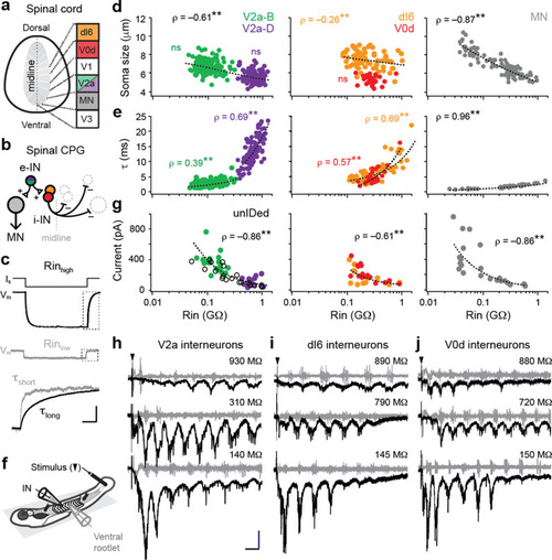
(a) Cross-section of spinal cord denoting canonical classes of molecularly defined locomotor-related interneurons. (b) Wiring diagram of swimming circuitry comprised of excitatory interneurons (e-IN) that provide local excitation (+) of motor neurons (MNs) and inhibitory (i-IN) interneurons, which cross the midline and silence neurons on the opposite side (?). Corresponding interneuron classes color coded as in Figure 1a. (c) Whole-cell current clamp recordings illustrate current step (Is) and membrane potential deflections (Vm) in high and low input resistance (R) dI6 interneurons. Expanded traces below from boxed regions are normalized to illustrate differences in time constant (?) related to resistance. Scale bar, 20 pA, 10 mV, 100 ms (top), and 10 ms (expanded). (d) Quantification of soma size versus input resistance for motor neurons and interneurons. Significant correlations are fit with logarithmic trendlines for illustrative purposes. ns, not significant. **, significant correlation following non-parametric Spearman Rank test. V2a-D=V2a neurons with descending axons; V2a-B=V2a neurons with bifurcating axons. V2a-B, ?(143)=?0.08, p=0.326, n=145; V2a-D, ?(103)=?0.14, p=0.219, n=105; dI6, ?(70)=?0.26, p<0.05, n=72; V0d, ?(26)=?0.09, p=0.641, n=28; MN, ?(104)=?0.87, p<0.001, n=106. Source data are reported in Figure 1?source data 1. (e) Quantification of membrane time constant versus input resistance. V2a-B, ?(152)=0.39, p<0.001, n=154; V2a-D, ?(103)=0.69, p<0.001, n=105; dI6, ?(68)=0.69, p<0.001, n=70; V0d, ?(26)=0.57, p<0.001, n=28; MN, ?(39)=0.96, p<0.001, n=41. Source data are reported in Figure 1?source data 1. (f) Schematic of the recording set up for ?fictive? swimming, evoked by a brief electrical stimulus to the tail skin (black triangle), with simultaneous recordings of interneuron (IN) activity and motor output from the ventral rootlet in chemically-immobilized larvae (see Materials and methods for details). (g) Quantification of peak inward excitatory currents versus input resistance during ?fictive? swimming.; V2a-B, ?(22)=?0.58, p<0.01, n=24; V2a-D, ?(12)=?0.11, p=0.702, n=14; V2a-unIDed, ?(17)=?0.92, p<0.001, n=19; V2a-pooled, ?(55)=?0.86, p<0.001, n=57; dI6, ?(11)=?0.44, p=0.133, n=13; V0d, ?(15)=?0.74, p<0.001, n=17; dI6-V0d-pooled, ?(28)=?0.61, p<0.001, n=30. Figure 1g has been adapted from Figure 2i from Kishore et al., 2014, distributed under the terms of a Creative Commons Attribution-Noncommercial-Share Alike 3.0 Unported License CC BY-NC-SA 3.0 (https://creativecommons.org/licenses/by-nc-sa/3.0/). It is not covered by the CC-BY 4.0 license and further reproduction of this panel would need to follow the terms of the CC BY-NC-SA 3.0 license. Source data are reported in Figure 1?source data 1. (h) Whole-cell voltage-clamp recordings of V2a interneurons at calculated chloride ion reversal potential (?65 mV) with simultaneous ventral rootlet recordings (gray) reveal rhythmic inward excitatory currents (black) driving ?fictive? swimming after a brief electrical stimulus to the skin (at black arrow). Input resistance values accompany respective traces. Scale bar, 50 pA, 25 ms. (i) As in (h) but for dI6 interneurons. (j) As in (h) but for V0d interneurons. © 2014, Kishore et al. All Rights Reserved. Figure 2i from Kishore et al., 2014, distributed under the terms of a Creative Commons Attribution-Noncommercial-Share Alike 3.0 Unported License CC BY-NC-SA 3.0 (https://creativecommons.org/licenses/by-nc-sa/3.0/). It is not covered by the CC-BY 4.0 license and further reproduction of this panel would need to follow the terms of the CC BY-NC-SA 3.0 license.
|

