
Fig. 2
- ID
- ZDB-FIG-220923-33
- Publication
- Kidokoro et al., 2022 - Nodal signaling regulates asymmetric cellular behaviors, driving clockwise rotation of the heart tube in zebrafish
- Other Figures
- All Figure Page
- Back to All Figure Page
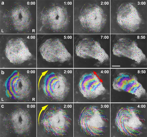
a Selected images from a confocal time-lapse recording of a Tg(myl7:EGFP-CAAX)ncv536Tg embryo starting at 20 hpf (Supplementary Movie 1). Cell membranes of myocardial cells are specifically labeled with GFP in the transgenic line. Dorsal view (anterior to the top). Relative times after the initiation of the recording (h:min) are indicated in the upper right corner. Color dots indicate tracking of individual myocardial cells. Images are representative of n ≥ 10 embryos. Scale bar = 50 µm. b Annotated duplicate images. Cells in the left primordium are colored. The group of cells converged in the circumferential direction toward the anterior seam of the left (L) and right (R) primordia (yellow arrow in b), while extending perpendicularly (red arrow in b) as the disc transformed into a tube. c Duplicated images overlaid with cell trajectories. Cell rearrangement and cell shortening occurred circumferentially toward the anterior seam of the left and right primordia (yellow arrow in c). |