|
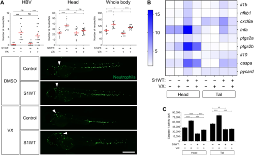
Fig. 2. WT S1 signals through the canonical inflammasome in zebrafish. Recombinant S1WT or vehicle (−) were injected in the hindbrain ventricle (arrowheads) of 2-dpf Tg(mpx:eGFP) (A) and WT (B and C) larvae in the presence of either dimethyl sulfoxide (DMSO) or the caspase-1 inhibitor VX-765 (VX). Neutrophil recruitment and number were analyzed at 3 hpi by fluorescence microscopy (A), the transcript levels of the indicated genes were analyzed at 12 hpi by RT-qPCR (B), and caspase-1 activity was determined at 24 hpi using a fluorogenic substrate (C). Representative images of whole Tg(mpx:eGFP) larvae for each treatment are also shown (A). Each dot represents one individual and the means ± SEM for each group is also shown. RT-qPCR data are depicted as a heatmap in (B) with higher expression shown in darker color. P values were calculated using one-way ANOVA and Tukey multiple range test. *P ≤ 0.05, **P ≤ 0.01, and ***P ≤ 0.001. Scale bar, 500 μm.
|