|
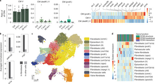
Cell type diversity of cardiac fibroblasts.a, Left: relative changes in abundance of different subtypes of CMs across the time points (n = 3, 9, 9, 5 animals; error bars show s.e.m.). Right: differentially expressed genes between subtypes of CMs. b, Comparison of average normalized expression of known proregenerative factors in fibroblasts and other cell types. The comparison used data pooled over all time points, and fibroblasts were defined as all cells in the yellow cluster in Fig. 1b. c, UMAP representation of the subclustering of col1a1a-expressing cells. d, Expression of ECM-related genes in different fibroblast cell types. The genes were classified according to their contribution to structure, breakdown or interaction of the ECM. A, atrium; Ctrl., control; const., constitutive; dediff., dedifferentiated; prolif., proliferating; V, ventricle. Source data
|