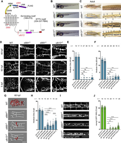
Figure 2. Germline mutants of the zebrafish Gpr124 ICD exhibit brain vascular defects
(A) Scheme of zebrafish Gpr124 highlighting the TALEN or CRISPR-Cas9 target sites corresponding to the gpr124s984, gpr124s985, and gpr124ulb13 alleles.
(B) Lateral views of WT, gpr124s984/s984 (s984−/−), gpr124s985/s985 (s985−/−), and gpr124ulb13/ulb13 (ulb13−/−) embryos at 60 hpf.
(C) Lateral views of the adult WT and gpr124 mutant skin pigmentation patterns. Arrowheads point at stripe discontinuities.
(D) Dorsal views of hindbrain vasculaturesgpr124 mutants.
(E) Quantification of hindbrain CtAs of WT and gpr124 mutant embryos at 48 hpf.
(F) Same as (E) at 60 hpf.
(G) 3D representation of the cerebrovasculature of 60 hpf WT and gpr124 mutant embryos. Red vessels are Wnt7a/Gpr124/Reck-dependent CtAs that sprout from white perineural vessels.
(H) Same as (E) in 5 dpf larvae.
(I and J) Dorsal views (I) and quantification (J) of dorsal root ganglia in the trunk region of WT and gpr124 mutant larvae at 72 hpf.
Scale bars, 0.5 mm for (B) and 100 μm for (D), (G), and (I). Related to Figure S1.