
FIGURE
Fig. 2
- ID
- ZDB-FIG-220504-17
- Publication
- Meng et al., 2021 - Cardiac toxicity assessment of pendimethalin in zebrafish embryos
- Other Figures
- All Figure Page
- Back to All Figure Page
Fig. 2
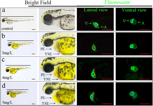
Heart morphology of zebrafish embryos exposed to PND for 72 h. With the increase of exposure concentration, pericardial and yolk sac edema increased (Bright Field), the SV-BA distance was increased (Fluorescent, Lateral view), and cardiac looped incomplete (Fluorescent, Ventral view). Green arrow indicates the atria and ventricles, Red line represents the SV-BA distance. PE, pericardial edema; YSE, yolk sac edema; SV-BA, sinus venous and bulbus arteriosus; A, atrium; V, ventricle; Scale = 350 µm. |
Expression Data
Expression Detail
Antibody Labeling
Phenotype Data
| Fish: | |
|---|---|
| Condition: | |
| Observed In: | |
| Stage: | Protruding-mouth |
Phenotype Detail
Acknowledgments
This image is the copyrighted work of the attributed author or publisher, and
ZFIN has permission only to display this image to its users.
Additional permissions should be obtained from the applicable author or publisher of the image.
Full text @ Ecotoxicol. Environ. Saf.