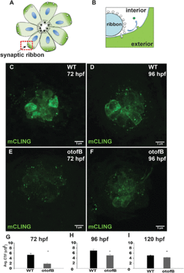
The otofB mutation results in reduced mCLING uptake. (A) Diagram of a neuromast in the same top-down orientation as the images in panels C–F. (B) Magnified view of a ribbon synapse highlighted in red in panel A. Endocytosis results in mCLING incorporation from the cell exterior into the vesicle lumen (green). (C, D) Representative confocal images of (C) 72 hpf and (D) 96 hpf mCLING-stained wild-type lateral line neuromasts. (E, F) Representative confocal images of (E) 72 hpf and (F) 96 hpf mCLING-stained mutant lateral line neuromasts. (G) Quantification of average mCLING dye associated with 72 hpf neuromasts of wild-type and otofB zebrafish (t test, p < 0.001). (H) Quantification of average mCLING dye associated with 96 hpf neuromasts of wild-type and otofB zebrafish (t test, p < 0.001). N = 6 larvae, 4 neuromasts per larvae for both wild type and mutant. Scale bars = 5 µm. (I) Quantification of average mCLING dye associated with 120 hpf neuromasts of wild-type and otofB zebrafish (t test, p < 0.001). N = 4 WT larvae, 3 mutant larvae, 4 neuromasts per larvae. Scale bars = 5 µm.
|
