|
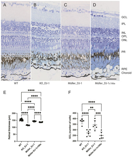
Laminar morphology of wild-type, knockout and transgenic retinas. Light microscopic images of toluidine-blue-stained retinal cross-sections from nine-month-old adult zebrafish: (A) wild type, (B) DJ-1 knockout, (C) Müller-cell-expressed wild-type DJ-1, (D) Müller-cell-expressed DJ-1c106a mutant. (E). Retinal thickness (μm) measured on eye sections from three fish in each group. **** p < 0.0001, one-way ANOVA, n = 17 (WT), 10 (KO_DJ-1), 15 (Müller_DJ-1) and 19 (Müller_DJ-1c106a). (F) Number of cells in ganglion cell layer measured on sections from three fish; ** p < 0.01, *** p < 0.001 and **** p < 0.0001 versus wild type, one-way ANOVA, n = 6). GCL, ganglion cell layer; IPL, inner plexiform layer; INL, inner nuclear layer; OPL, outer nuclear layer; PR, photoreceptors; RPE, retinal pigment epithelium; V, vacuole in RPE layer. Bar, 20 μm applies to all panels.
|